
|
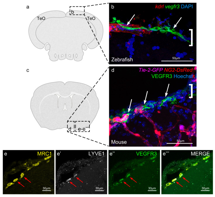
Figure 3 Murine leptomeninges contain cells expressing BLEC markers. Image adapted from Shibata et al. [55]. (a) and (c) Coronal sections of a zebrafish and a mouse brain, respectively, equivalent to the imaging areas in (b,d). To facilitate comparison, (b,d) are both displayed with the parenchyma at the bottom and the meninges at the top. (b) Zebrafish BLECs (white arrows) express Vegfr3 (green) and are located adjacent to a meningeal blood vessel (red). The area of the meninges is marked with a white bracket. DAPI (blue) highlights the nuclei. (c) IHC of a 17 week old mouse reveals the presence of VEGFR3 expressing cells (white arrows) adjacent to meningeal blood vessels (magenta). These cells are limited to the region of the meninges (white bracket) and do not penetrate into the brain parenchyma. NG2 (red) labels pericytes and smooth muscle cells, Hoechst (blue) marks nuclei. (e–e’’’) IHC of mouse brain section showing that the cells within the meningeal layers express the BLEC markers MRC1, LYVE1 and VEGFR3.