
|
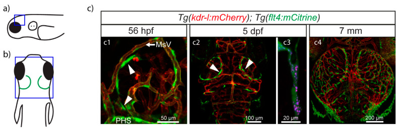
Figure 2 Zebrafish BLECs in embryonic and larval specimens. (a,b) Schematic representations of zebrafish showing the same orientation and close-up regions (blue squares) as in c1 (a) and c2-c4 (b). (c) Confocal images from van Lessen et al. [50]. Lympho-venous structures are depicted in green and blood vessels in red. BLECs (arrow heads) start sprouting at 56 hpf from behind the PHS and migrate along the MsV (c1). By 5 dpf they have formed a bilateral loop in the TeO (c2). Cells in the loop are able to take up injected dyes such as pHrodo (red) and IgG-Alexa647 (blue) (c3). BLECs keep spreading above the whole brain area (c4), where they will stay as single cells throughout the lifespan of the animal. Dpf, days post fertilization; hpf, hours post fertilization; MsV, mesencephalic vein; PHS, primary head sinus; TeO, optic tectum.