IMAGE

Figure 3

ID
ZDB-IMAGE-210430-3
Source
Figures for Villamayor et al., 2021
Image
Figure Caption

Figure 3

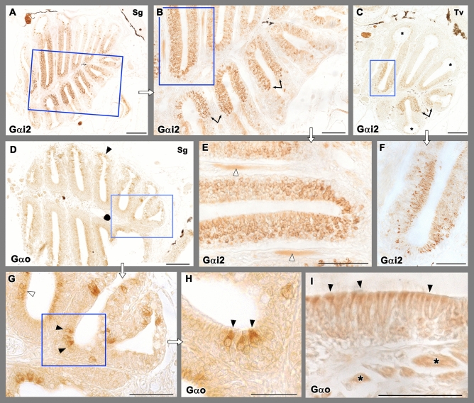
Immunohistochemical study of the olfactory rosette of zebrafish with antibodies against G-proteins. (AC,E,F) Anti-Gαi2 immunolabelling. (A) Sagittal sections show a higher number of immunopositive neurons in the internal parts of the rosette, at both sides of the median raphe. (B) Higher magnification of the inset in (A). The transition between both the nonsensory and sensory epithelium is straight (arrows). (C) Transverse sections show how the lateral rim of the lamella (asterisks) lacks of anti-Gαi2 immunolabelling. (E) Inset in (B) showing the immunopositive olfactory nerves (white arrowheads). (F) Inset in C shows how the basal part of the neuroepithelium lacks immunopositive cells. (D,GI) Anti-Gαo immunolabelling. (D) The immunoreactivity was present diffusely in apical neurons, as it is shown by open arrowheads in the inset (G). Additionally, more isolated big and oval receptor neurons showed immunoreactivity (black arrowheads) (H, inset in G). (I) The processes in the nonsensory epithelium (arrowheads) and the branches of the olfactory nerves (asterisks) are also immunopositive. Scale bar: 100 μm (A,C,D); 50 μm (B,EI).

Acknowledgments
This image is the copyrighted work of the attributed author or publisher, and ZFIN has permission only to display this image to its users. Additional permissions should be obtained from the applicable author or publisher of the image. Full text @ Sci. Rep.