
|
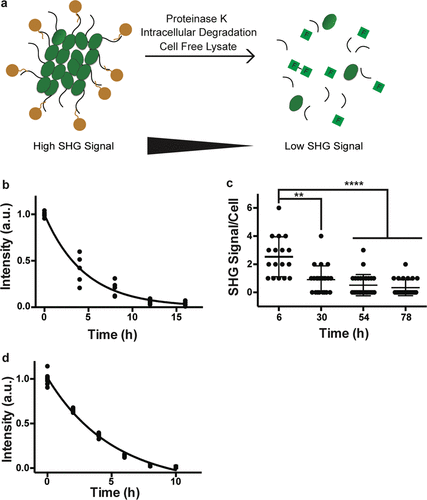
Fig. 3 Bioharmonophores can be degraded by proteases, cells, and cell-free lysate systems. (a) Schematic showing different degradation methods utilized to assess biodegradability of the bioharmonophores. (b) Graph displaying the change of SHG signal intensity over time of bioharmonophores incubating with proteinase K (n = 5). Mean values of data points were fitted for one-phase exponential decay. (c) Quantification of SHG signal/cell after overnight incubation of Tat-peptide functionalized bioharmonophores over time. SHG signal/cell is significantly reduced 30 h after reseeding. Mean ± s.d. ****, P < 0.0001, **, P < 0.005, *, P < 0.05 (nonparametric Kruskal–Wallis test with Dunn’s post hoc multiple comparison). (d) Graph showing the loss of SHG signal intensity when bioharmonophores are subjected to the cell-free reticulate lysate degradation system (n = 5). Mean values of data points were fitted using a one phase exponential decay. Scale bar, 10 μm (c); 10 μm (d).