IMAGE

Fig. 2

ID
ZDB-IMAGE-210419-6
Source
Figures for Don et al., 2021
Image
Figure Caption

Fig. 2

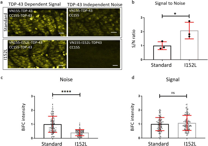
Optimized mVenus fragment (VN155-I152L) increases the signal-to-noise ratio of TDP-43 complementation. a Representative microscope images demonstrating the fluorescence reconstitution in muscle tissue at 24 hpf using VN155-TDP-43 versus VN155-I152L-TDP-43 constructs. Scale bars represent 20 μm. b Quantitative comparison of signal-to-noise ratio using standard VN155 versus VN155-I152L fragments. Data was obtained using an unbiased plate reader. Data pooled and averaged from three independent replicates. c, d The background fluorescence (noise) was significantly reduced with the I152L mutation (c), while the maximum fluorescence intensity did not change (d). Data in c, d was obtained using an unbiased plate reader. Data pooled from three independent replicates. *P < 0.05; ****P < 0.0001; nsP > 0.05

Acknowledgments
This image is the copyrighted work of the attributed author or publisher, and ZFIN has permission only to display this image to its users. Additional permissions should be obtained from the applicable author or publisher of the image. Full text @ Mol. Neurobiol.