
|
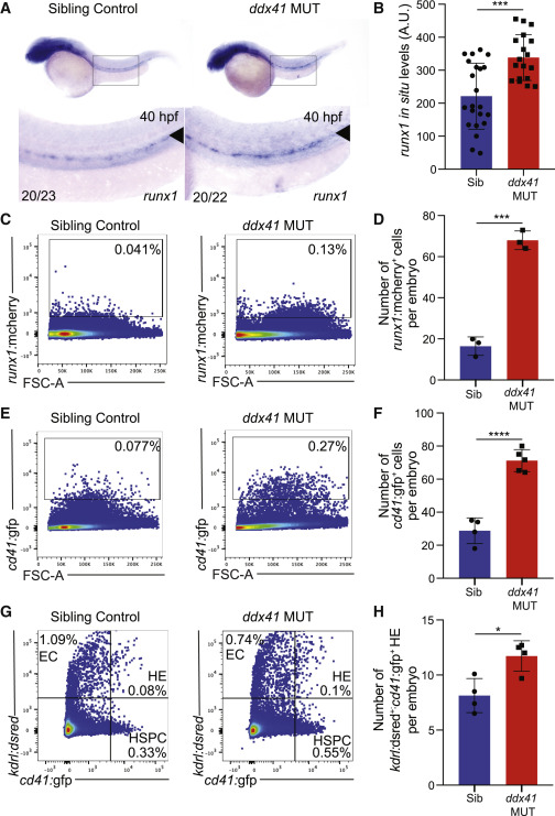
Fig. 1 Ddx41 regulates HSPC number (A) In situ hybridization of the HSPC marker runx1 at 40 hpf in sibling controls (left) and ddx41 mutants (right). Numbers on bottom left corner indicate the fraction of embryos with the same phenotype as the one depicted in the image. Inset underneath shows a higher magnification (8×) view of the boxed AGM region above. The aorta is marked with arrowheads. (B) Quantification of runx1 in situ hybridization levels from (A). Quantification was done using Fiji. a.u., arbitrary unit. (C and E) Flow cytometry plots of runx1:mcherry+ (C) and cd41:gfp+ (E) HSPCs from sibling controls (left) and ddx41 mutants (right) at 40 hpf. (D and F) Graphs depicting the absolute number of runx1:mcherry+ (D) and cd41:gfp+ (F) per embryo at 40 hpf. (G) Flow cytometry plots of kdrl:dsred+, cd41:gfp+, and kdrl:dsred+; cd41:gfp+ double positive cells from sibling controls (left) and ddx41 mutants (right) at 40 hpf. (H) Graphs depicting the absolute number of kdrl:dsred+; cd41:gfp+ double positive hemogenic endothelial cells per embryo at 40 hpf. Graphs display means ± standard deviations (stds) with p values calculated with unpaired Student’s t test, ∗p < 0.05, ∗∗∗p < 0.001, ∗∗∗∗p < 0.0001. N = 3–5 replicates per experiment.
Reprinted from Developmental Cell, 56(5), Weinreb, J.T., Ghazale, N., Pradhan, K., Gupta, V., Potts, K.S., Tricomi, B., Daniels, N.J., Padgett, R.A., De Oliveira, S., Verma, A., Bowman, T.V., Excessive R-loops trigger an inflammatory cascade leading to increased HSPC production, 627-640.e5, Copyright (2021) with permission from Elsevier. Full text @ Dev. Cell