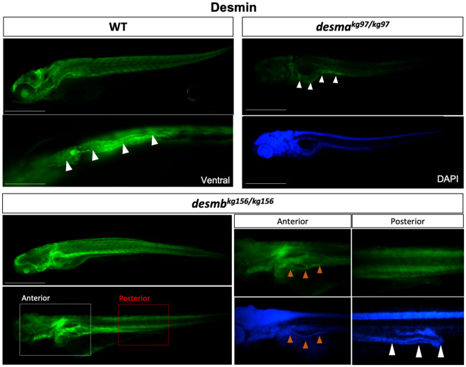
IMAGE

Figure 3

ID
ZDB-IMAGE-210409-4
Antibodies
Source
Figures for Kayman Kürekçi et al., 2021
Image
Figure Caption

Figure 3

Desmin protein expression in 96 hpf embryos. Whole mount immunofluorescence staining of desmin with anti-desmin polyclonal antibody recognizing both Desma and Desmb, in wild-type and mutant 96 hpf embryos. Left upper panel is lateral view of a WT embryo with anterior to left, dorsal to top, beneath is ventral view with anterior to left, white arrowheads indicate the gut. Right upper panels are lateral views of desmakg97/kg97 with anterior to left, dorsal to top. Lower panel shows desmbkg156/kg156 embryos with anterior to left, dorsal to top. Scale bar: 500 µm. Boxes with dashed lines delineate areas that were zoomed and focused on anterior and posterior segments of the intestine as shown in lower right panels, orange arrowheads indicate anterior intestine, white arrowheads show middle and posterior segments of the intestine.

Figure Data
Acknowledgments
This image is the copyrighted work of the attributed author or publisher, and ZFIN has permission only to display this image to its users. Additional permissions should be obtained from the applicable author or publisher of the image. Full text @ Sci. Rep.