
|
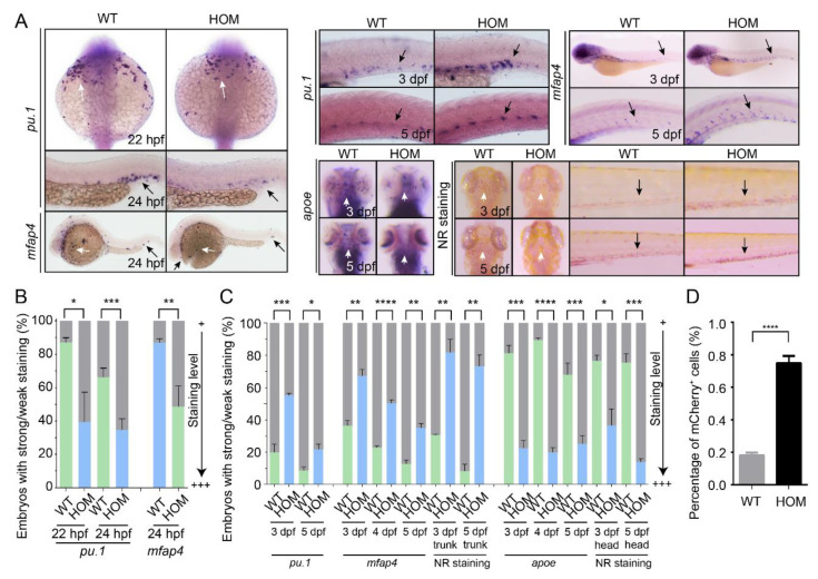
Figure 4 Macrophage development in WT and mafbb mutant embryos. (A) WISH of pu.1, mfap4 and apoe in embryos at 22–24 hpf. WISH of pu.1, mfap4 and apoe, neutral red (NR) staining in embryos at 3–5 dpf. The white arrows point to the rostral blood island and yolk sac, the black arrows point the ventral tail region. (B) Quantification of WISH results in embryos at 22–24 hpf (n = 50–100 embryos per group). (C) Quantification of WISH and NR staining in embryos at 3–5 dpf (n = 50–100 embryos per group). (D) Quantification of mCherry+ macrophage cells from Tg(mpeg1:mCherry) embryos at 5 dpf by flow cytometry. WT, wild type; HOM, mafbbd11/d11. Results in B-D are expressed as mean ± SEM, (* p < 0.05, ** p < 0.01, *** p < 0.001, **** p < 0.0001, t test).