
|
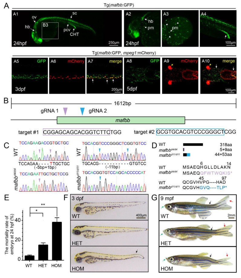
Figure 1 The expression of mafbb during embryogenesis and generation of mafbb mutants. (A) Tg (mafbb:GFP) shows mafbb expression in primitive myeloid (pm) cells, hindbrain (hb), otic vesicle (ov), spinal cord (sc), posterior cardinal vein (pcv), caudal hematopoietic tail region (CHT) (A1–4). A2–4 are enlarged images of A1. The white arrowheads in A2 and A3 point to pm cells. The double Tg(mafbb:GFP;mpeg1:mCherry) shows the expression of GFP in mCherry+-macrophages in the CHT (A5–7, lateral views) and in the head region (A8–10, ventral views). The white arrows in (A7 and A10) point to the overlapped expression of mafbb and mepg1. The arrowheads in (A9–10) mark the eyes from α-crystallin-mCherry in the plasmid of mabb:GFP [32]. (B) The schematic diagram of mafbb cDNA and the targeted regions of two guide RNAs. The target DNA sequences are shown in purple or blue rectangles. (C) Sanger sequencing analysis of PCR fragments containing gRNA1 and gRNA2 targeted regions from mafbb deficient homozygotes. The deleted nucleotides are replaced by -, and the inserted nucleotides are in red. (D) Schematic representation and amino acid sequence of the wild type MafBb and two predicted truncated proteins. (E) The mortality rate of embryos at 24 hpf (n = 315–688 per group). Results are expressed as mean ± SEM, (* p < 0.05, ** p < 0.01, t test). The statistical significance was displayed as (F) Images of embryos at 3 dpf. The black arrow points to the curved tail. (G) Images of adult zebrafish at 9 mpf. The projecting lower jaw (green arrows), the curved spine (black arrows) and the asymmetric caudal fin (red arrows) are shown in mafbb−/− mutants. WT, wild type; HET, mafbbd11/+; HOM, mafbbd11/d11; mpf, month post fertilization.