IMAGE

Figure 6

ID
ZDB-IMAGE-210409-11
Source
Figures for Kayman Kürekçi et al., 2021
Image
Figure Caption

Figure 6

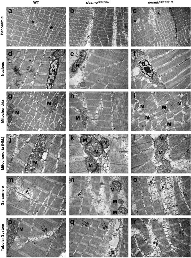
Ultrastructural features of adult WT and mutant skeletal muscle tissue. First, second and third columns present adult WT, desmakg97/kg97, desmbkg156/kg156 skeletal muscle tissues’ electron micrographs, respectively. First row indicates the generally aligned sarcomeric structures with the presence of seldom disruptions (ac, asterisks). Second row shows centrally (d) and peripherally (e,f) located healthy nuclei (N) with peripherally condensed chromatin and well-defined nucleoli. Third and fourth row (g–l) shows pleomorphic mitochondria (M) with well-defined tubular cristae. Fifth and sixth rows present the Z line streaming (arrows in mo) and enlargement (mr) of the T-tubule systems related to the triads (double arrows). Uranyl acetate, lead citrate. HM high magnification. Magnifications are as follows: (ac) 8000 ×, (d) 25,000 ×, (e,f) 20,000 ×, (gi) 15,000 ×, (j) 40,000 ×, (k) 60,000 ×, (l) 30,000 ×, (mo) 20,000 ×, (p) 25,000 × and (q,r) 20,000 ×.

Figure Data
Acknowledgments
This image is the copyrighted work of the attributed author or publisher, and ZFIN has permission only to display this image to its users. Additional permissions should be obtained from the applicable author or publisher of the image. Full text @ Sci. Rep.