
|
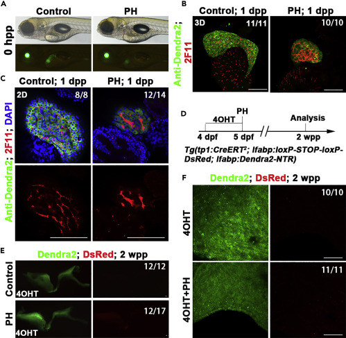
Fig. 5 BECs fail to contribute to hepatocyte regeneration after PH (A) Live images showing the liver (Green) after PH at 0 hpp. (B) Confocal projection showing (3D imaging) the co-immunostaining for Dendra2 and 2F11 at 1 dpp. (C) Single optical images showing the co-immunostaining for Dendra2 and 2F11 at 1 dpp. The 2F11+ (BECs) were Dnedra2-. Nuclei were stained with DAPI (blue). (D) Experimental scheme illustrating the stage of 4OHT and PH and analysis at 2 wpp in triple transgenic line Tg(tp1:CreERT2; lfabp:loxP-STOP-loxP-DsRed; lfabp:Dendra2-NTR). (E) The confocal projection shows that no DsRed expression in regenerating livers at 2 wpp. (F) The confocal projection (3D imaging) shows that no DsRed expression at 2 wpp in large magnification. Numbers indicate the proportion of larvae exhibiting the expression shown. Scale bars: 100 μm. hpp: hours post-partial hepatectomy; dpp: days post-partial hepatectomy; wpp: weeks post-partial hepatectomy.