
|
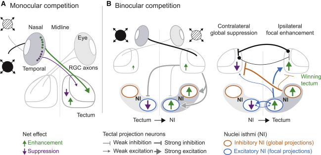
Fig. 8 (A) Schematized summary of findings for monocular competition. The most salient stimulus “wins” in the retina through reciprocal inhibition, possibly mediated by amacrine cells. Saliency tuning is sharpened by a tectum-intrinsic circuit. (B) Circuit model for binocular competition. Each tectum drives activity in the ipsilateral NI (putative excitatory tectobulbar neurons) and suppresses activity in the contralateral NI (putative inhibitory intertectal neurons). Focal enhancement, mediated by excitatory NI cells, is stronger on the “winning” stimulus side (green arrows). Suppression, mediated by globally projecting inhibitory NI neurons, is stronger on the “losing” stimulus side (magenta arrows). Reciprocal isthmotectal loops ensure focal enhancement of responses to a stronger stimulus and suppression of responses to a weaker stimulus, implementing a WTA computation. Black connections across both tecta represent putative inhibitory commissural neurons projecting to the contralateral side. Equal activity in both NIs may result in neither tectum winning and an averaging strategy being implemented instead.