
|
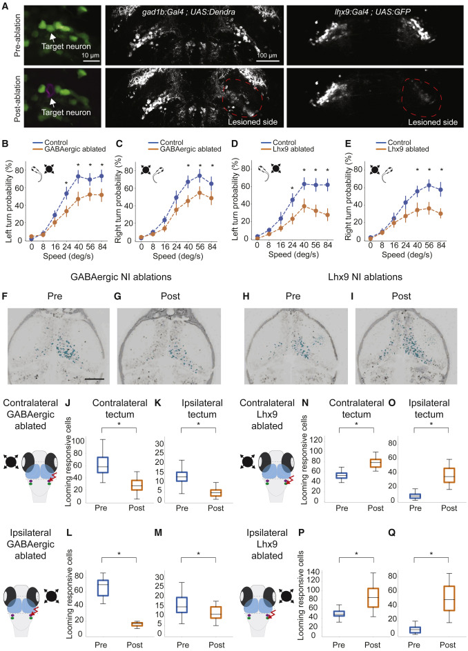
Fig. 6 (A) 2P laser ablation of isthmic neurons. Shown is an example of single cell ablation (left panels). After ablation of a cell, red fluorescence (magenta) is visible in the target spot. The center panels show representative images of unilateral 2P laser ablation of GABAergic-positive isthmic neurons in gad1b:Gal4VP16mpn155; UAS:Dendra-krass1998t pre-ablation and post-ablation. Right panels: ablation of glutamatergic isthmic neurons in lhx9:Gal4VP16mpn203; UAS:EGFP pre-ablation and post-ablation. (B) Probability of left escapes in control (blue) and GABAergic NI-ablated fish (orange). (C) Similar to (B) but for right escapes. (D) Probability of left escapes in control (blue) and lhx9 NI-ablated fish (orange). (E) Similar to (D) but for right escapes. For all looming-evoked escape panels, error bars represent SD. n = 12 for control fish (blue). n = 15 for ablated fish (orange). ∗p < 0.05, Tukey’s honestly significant difference (HSD) test. (F and G) Example of the effect of unilateral ablation of the GABAergic-positive NI on the right hemisphere. (F) is before ablation and (G) is after ablation. The corresponding t-statistic for each pixel is calculated for looming-responsive cells and labeled in cyan. (H and I) Example of the effect of unilateral ablation of the lhx9-positive NI on the right hemisphere before (H) and after (I) ablation. (J) Number of looming-responsive cells in the contralateral (relative to looming stimulus) tectum before and after ablation of GABAergic-positive NI cells in the right hemisphere. n = 4. (K) Number of looming-responsive cells in the ipsilateral (relative to looming stimulus) tectum. (L) Number of looming responsive cells in the contralateral tectum (relative to looming stimulus) for the intact hemisphere before and after ablation of GABAergic-positive NI cells in the right hemisphere. N = 2. (M) Number of looming-responsive cells in the ipsilateral tectum (relative to looming stimulus) for the intact hemisphere after ablation of the GABAergic-positive NI. (N) Number of looming-responsive cells in the contralateral (relative to looming stimulus) tectum before and after ablation of lhx9-positive NI cells in the right hemisphere. n = 3 fish. (O) Number of looming-responsive cells in the ipsilateral (relative to looming stimulus) tectum. (P) Number of looming-responsive cells in the contralateral tectum (relative to looming stimulus) for the intact hemisphere before and after ablation of lhx9-positive NI cells in the right hemisphere. n = 2 fish. (Q) Number of looming-responsive cells in the ipsilateral tectum (relative to looming stimulus) for the intact hemisphere after ablation of lhx9-positive NI cells. For all panels, ∗p < 0.05, Mann-Whitney U test.