
|
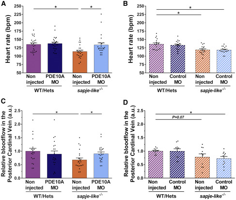
Fig. 6 Improvement in Cardiovascular Activity of pde10a Morphant Dystrophin-Deficient Zebrafish Pairs of sapje-like+/− zebrafish were mated and pde10a morpholino or standard control morpholino (negative control) was injected in one-cell-stage progeny embryos. At 4 dpf, a birefringence assay was performed and unaffected larva (normal muscle birefringence phenotype) as well as affected larvae (abnormal muscle birefringence phenotype) were sorted. At 5 dpf, anesthetized larvae were recorded for at least 30 s under a microscope. In each experiment, morphant zebrafish were compared to non-injected siblings. At the end of the experiment, fish were collected and genotyped. (A and B) Dot plots show the heart rate (beats per minute [bpm]) of 5-dpf larvae in (A) pde10a morpholino experiments and (B) control morpholino experiments. Seventeen to 24 (n = 17–24) zebrafish were analyzed within at least three (N = 3) independent experiments. Statistical differences between groups are presented as follows: ∗p < 0.05 (t test, ±SEM). (C and D) Dot plots show blood flow (arbitrary units [a.u.]) of posterior cardinal vein in (C) pde10a morpholino experiments and (D) control morpholino experiments. Blood flow was measured by using DanioScope software (Noldus). Ten to 22 (n = 10–22) zebrafish were analyzed within at least three (N = 3) independent experiments. Statistical differences between groups are presented as follows: ∗p < 0.05 (t test, ±SEM).