
|
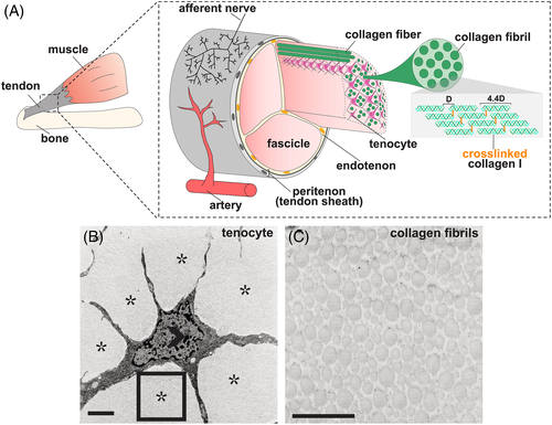
Fig. 1 Tendon structure. A, Graphical representation of tendon morphology. The tendon midsubstance is comprised of collagen molecules which are spaced apart at a distance of 67 nm (letter D in diagram) and cross‐linked to form stable fibrils.15 Tenocytes are interspersed between collagen fibrils, which together generally form higher order bundles called fascicles. Fascicles are held together by connective tissue called the endotenon. The tendon midsubstance is encased in the peritenon, or tendon sheath, which is comprised of a basement membrane and epithelial cell layer.16 B,C, Transmission electron micrograph (TEM) of 6‐week‐old mouse tenocyte, B, and collagen fibrils, C. Open arrowhead marks cell nuclei and asterisks mark collagen fibrils. Rectangle in, B, is shown at higher magnification in, C. Scale bar, 1 μm. Images in, B,C, were adapted from Kalson et al17