
|
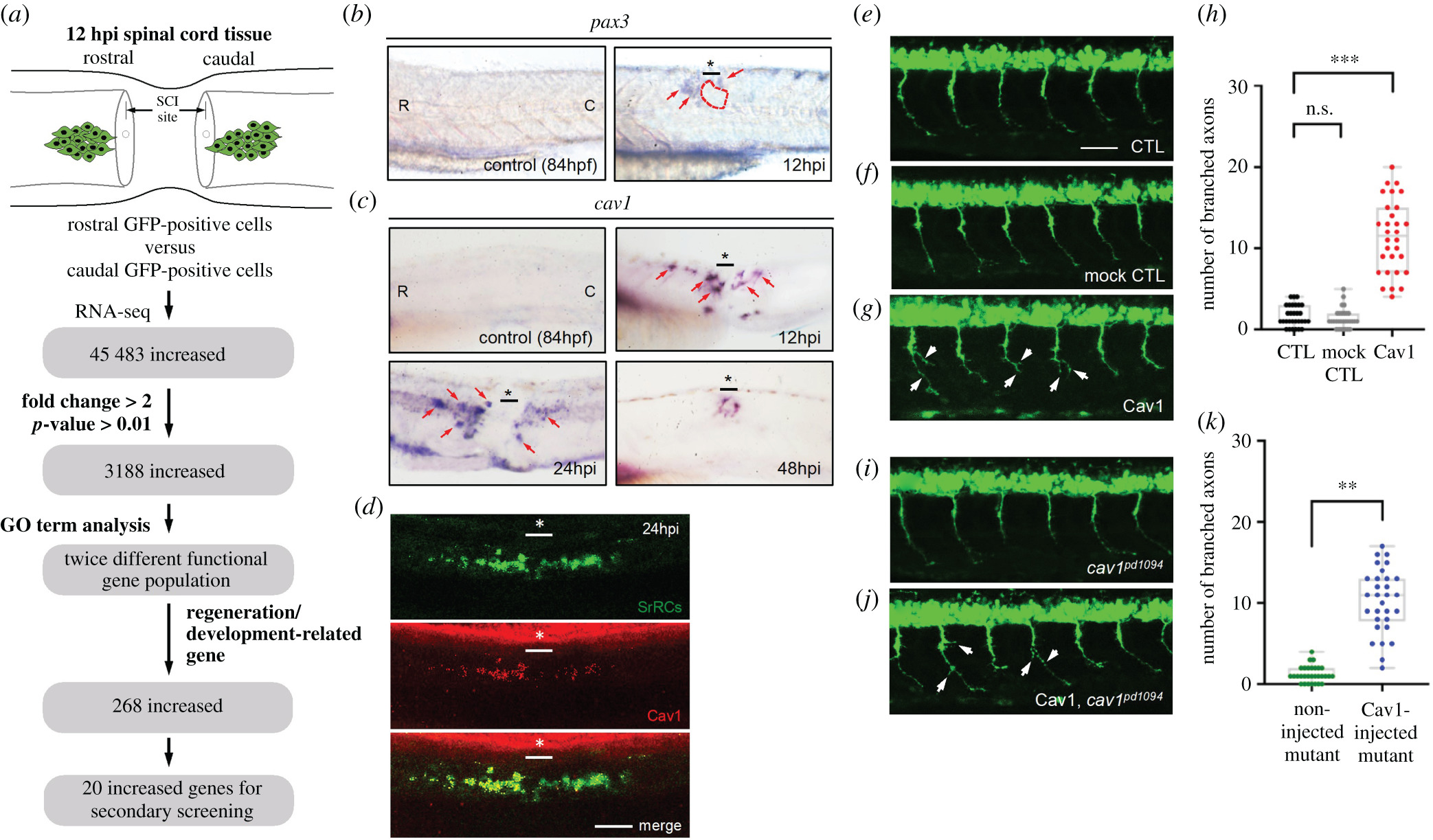
Fig. 6 Expression of cav1 transcript and encoded protein in zebrafish embryos. (a) Schematic illustration showing how putative genes were selected for this study. Using whole-mount in situ hybridization to detect (b) pax3 and (c) cav1 mRNAs in the spinal cord of zebrafish embryos before SCI (served as a negative control) and after SCI at 12, 24 and 48 h post-injury (hpi), as indicated. Since the expression pattern of pax3a, another transcript screened from RNA-seq, also showed increased expression in rostral-SrRCs compared to that in caudal-SrRCs, it served as a positive control in a parallel experiment. (d) Using IHC detection to examine the protein level of Cav1 (labelled by red signal) expressed in SrRCs (labelled by green signal), including rostral-SrRCs and caudal-SrRCs, in SCI-embryos at 24 hpi. The underscore stars indicate the SCI sites. Scale bar shown at lower right corner is 50 μm. (e) CaP motor neuron labelled by GFP was observed in the spinal cord of zebrafish embryos from transgenic line Tg(mnx:GFP) at 24 hpf (un-injected control. Student's t-test was used to perform statistical analysis, **p < 0.01 significance. CTL), (f) pCS2-vector-injected embryos (mock control) and (g) pCS2-Cav1-injected embryos. (h) Quantification and comparison of the number of branched axons of CaP motor neurons between groups. (i) GFP-labelled CaP motor neurons observed in the spinal cord of embryos from cav1 mutant (cav1pd1094) at 24 hpf (un-injected control). Student's t-test was used to perform statistical analysis, **p < 0.01 significance. (j) pCS2-Cav1-injected embryos from a double-transgenic line, in which mutant cav1pd1094 was crossed with Tg(mnx:GFP). White arrows indicate branched axons. (k) Quantification and comparison of the number of branched axons of CaP motor neurons between groups as indicated. Each spot indicates the number of axonal branches in each embryo (n = 30), in which six CaP motor neurons were analysed. Student's t-test was used to perform statistical analysis, ***p < 0.001 significance.