
|
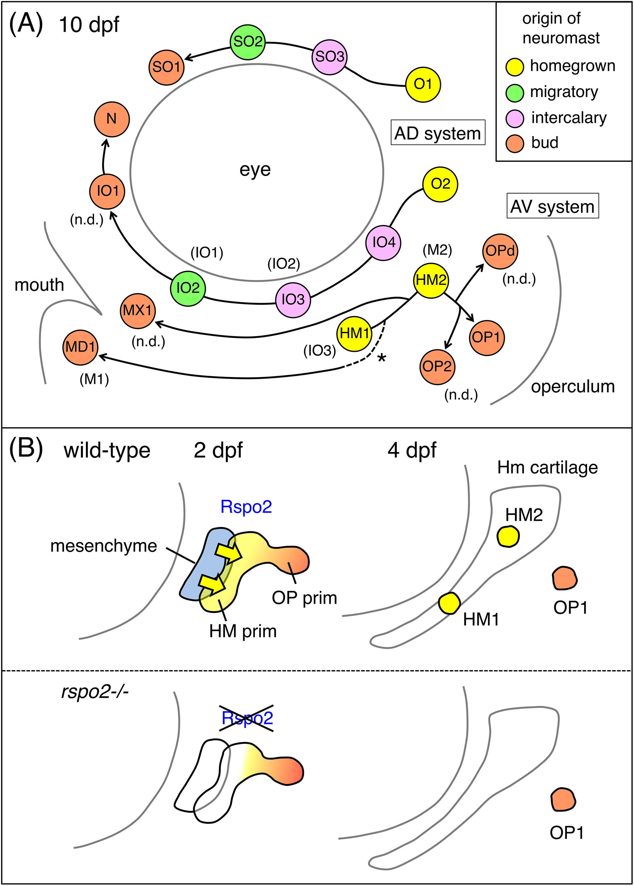
Fig. 8 Model for the development of the ALL neuromasts in zebrafish. A, Schematic representation for the ALL neuromasts at 10 dpf. The origin of each neuromast is indicated by a different color. The formation of MD1 by budding is presumptive (dotted line with an asterisk). Designations by Raible and Kruse14 are indicated in parentheses. “n.d.” indicates the neuromasts not described in the previous study. B, Model for the role of Rspo2 during the neuromast formation. Rpso2 (yellow arrows), emanated by the hyoid mesenchyme (blue), acts on the adjacent HM prim to promote proliferation and differentiation of HM1 and HM2 (yellow). Budding of OP1 (orange) from the HM prim takes place normally