
|
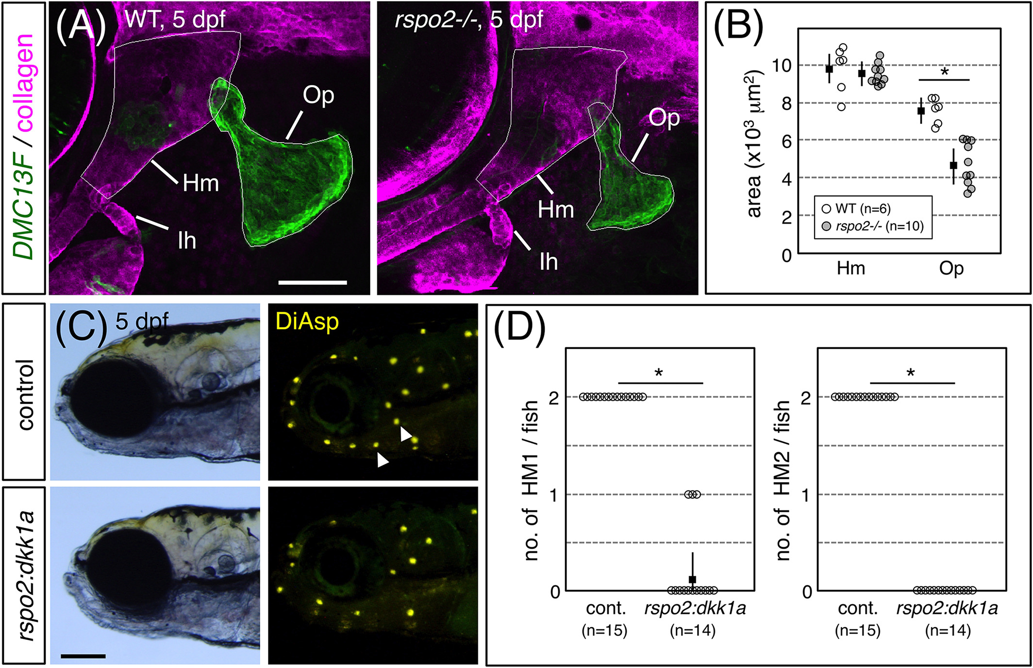
Fig. 7 Hyoid‐derived Rspo2 is required for the development of HM1 and HM2. A, 5 dpf‐DMC13F;UAS:gfp embryos, which express GFP in osteoblasts, stained with anti‐type II collagen antibody. Op, opercle. B, Quantification of sizes of Hm and Op defined by the outlines indicated in A. C, 5 dpf‐DMC131A(rspo2:gal4);UAS:dkk1a‐rfp and wild‐type sibling embryos stained with DiAsp. Note the specific loss of HM1 and HM2 (arrowheads). D, Quantification of numbers of HM1 and HM2 in wild‐type and dkk1a‐expressing embryos. Lateral views, anterior is to the left. Data are given as the mean ± SD (B, D) *P < .01 (t‐test). Scale bars: 100 μm (C); 50 μm (A)