
|
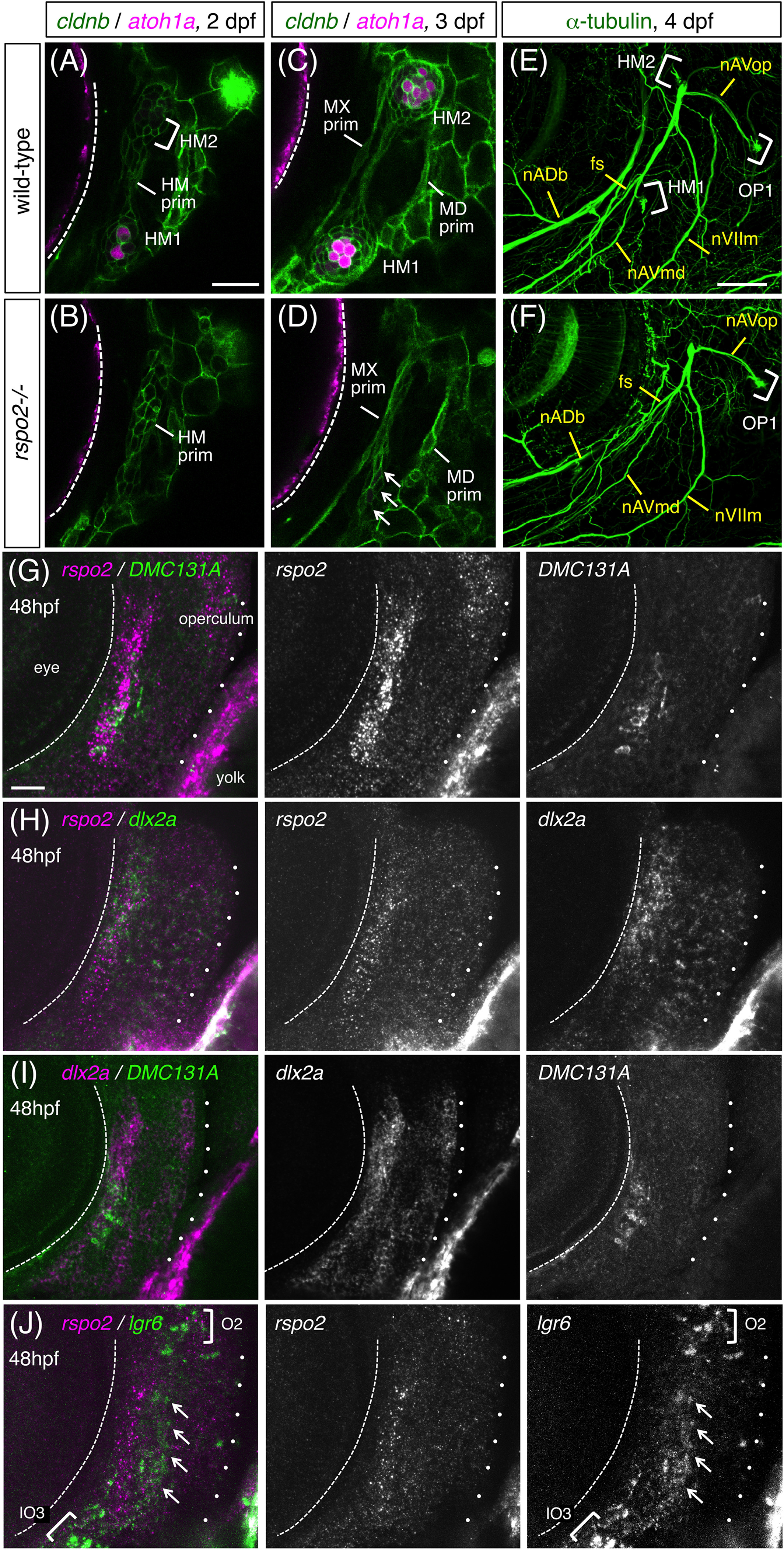
Fig. 6 Expression of rspo2 in hyoid mesenchyme. A‐D, Time‐course observations of wild‐type (A, C) and rspo2−/− (B, D) embryos with cldnb:gfp;atoh1a:rfp background at 2 dpf (A, B) and 3 dpf (C, D). Putative remnants of the HM prim are indicated by arrows in D. E, F, 4 dpf‐wild‐type (E) and rspo2−/− (F) embryos labeled with anti‐acetylated α‐tubulin antibody. fs, facial sensory nerve; nVIIm, facial motor nerve. G, 48 hpf‐DMC131A(rspo2:gal4);UAS:gfp embryo doubly labeled with anti‐GFP antibody and RNA probe for rspo2. H, 48 hpf‐embryo doubly labeled with RNA probes for rspo2 and dlx2a. I, 48 hpf‐DMC131A(rspo2:gal4);UAS:gfp embryo doubly labeled with anti‐GFP antibody and RNA probe for dlx2a. J, 48 hpf‐embryo doubly labeled with RNA probes for rspo2 and lgr6. The posterior margin of the eye is indicated by the dotted line. The operculum is outlined by dots. Lgr6‐expressing cells are indicated by arrows (J). Lateral views, anterior is to the left. Scale bars: 50 μm (E, F); 20 μm (A‐D, G‐J)