
|
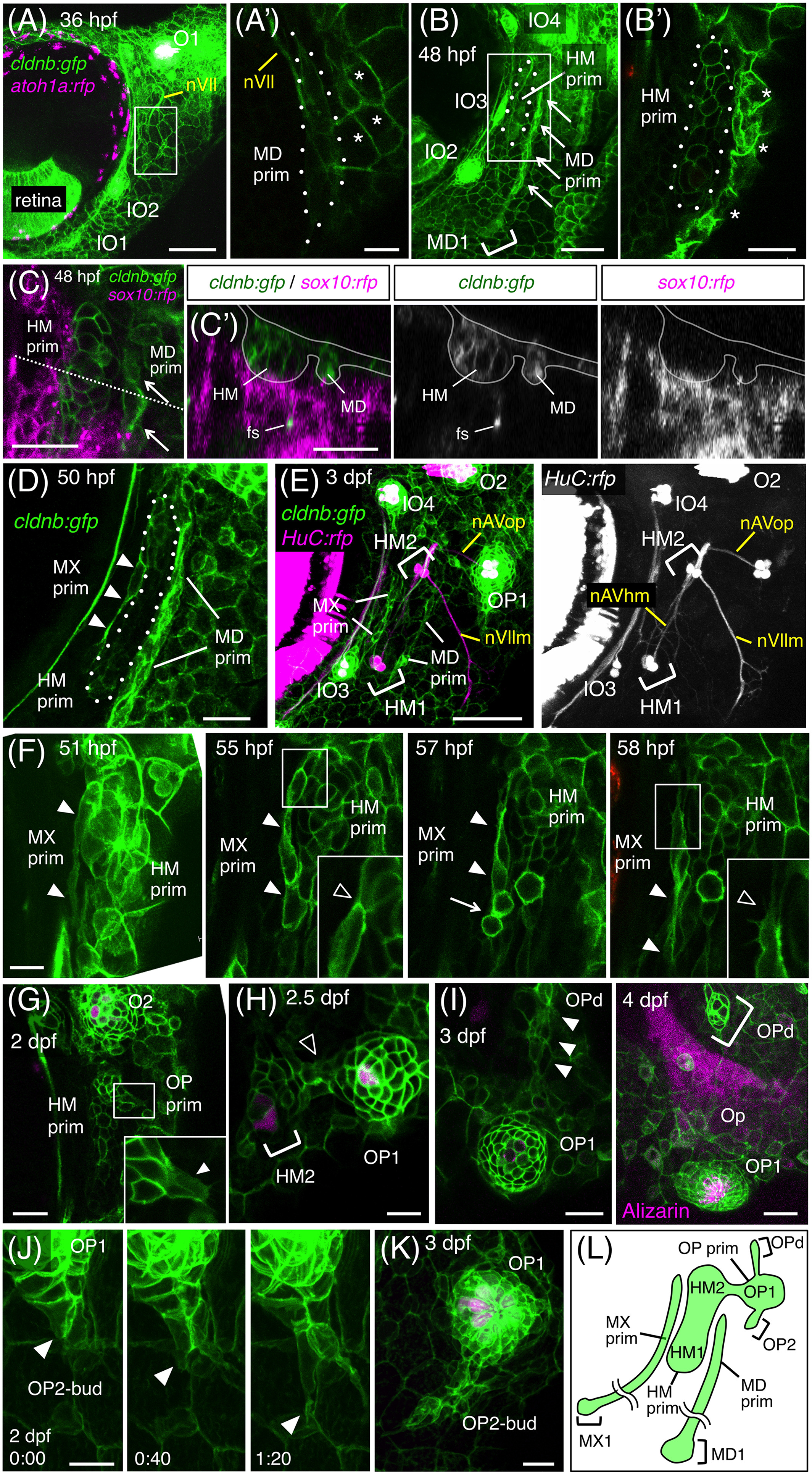
Fig. 4 Development of the anteroventral (AV) system. A, Hyoid region of cldnb:gfp;atoh1a:rfp embryo at 36 hpf. A′, Single confocal plane of the boxed area, showing a cluster of elongated cells of the MD prim (dotted line). nVII, facial nerve. Asterisks indicate cldnb:gfp‐expressing peridermal skin cells. B, Elongation of the HM prim at 48 hpf. MD1 forms in the ventral end of the MD prim. B′, Single confocal plane of the boxed area in B. C, Hyoid region of cldnb:gfp;sox10:rfp embryo at 48 hpf. C′, Optical cross‐section along the dashed line in C. fs, facial sensory nerve. D, Formation of the MX prim at 50 hpf. E, Differentiation of HM1 and HM2 in cldnb:gfp;HuC:gal4;UAS:rfp embryo at 3 dpf. F, Budding of the MX prim (indicated by arrowheads) from the HM prim. A single cell elongates (51 hpf), undergoes cell divisions (57 hpf, arrow), and extends ventrally (58 hpf). Insets indicate higher magnifications of the boxed areas, showing detachment of the MX cell from the HM prim. G, Budding of the OP prim from the HM prim at 36 hpf. Inset indicates a higher magnification of the boxed area, showing the budding cell (arrow). H, Differentiation of OP1 at 2.5 dpf. Cells connecting the HM prim and the OP prim are indicated by an open arrowhead. I, Time‐course observation of OPd formation at 3 and 4 dpf. The budding cell is indicated by arrows. Opercle bone (Op) is stained with Alizarin red. J, Time‐lapse observation of OP2 budding at 2 dpf. Arrowheads indicate the leading cell. K, Proliferation of OP2‐bud at 3 dpf. L, Schematic drawing for the developing AV system at 3 dpf. Lateral views, anterior is to the left. Scale bars: 50 μm (A, B, E, K); 20 μm (A′, B′, C, C′, D, G‐I); 10 μm (F, J)