
|
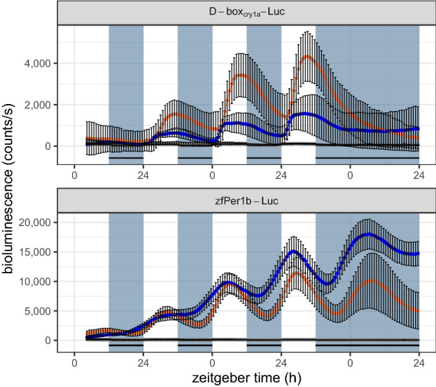
Figure 3 Luciferase assay showing rhythmic bioluminescence of Amphiprion ocellaris EAO (orange) and zebrafish PAC-2 cells (blue) transfected with D-boxCry1a-Luc (above) or zfPer1b-Luc (below) constructs. A negative control of non-transfected cells is shown in grey. For each condition, the mean of eight independently transfected wells and standard deviation is shown. Cells were exposed to a 12:12 h LD cycle, indicated by bluish-grey background and black stripes. Both cell lines, transfected with zfPer1b-Luc showed a comparable robust daily rhythm, even under DD. Cells transfected with D-boxCry1a-Luc were only rhythmic during the LD cycle.