
|
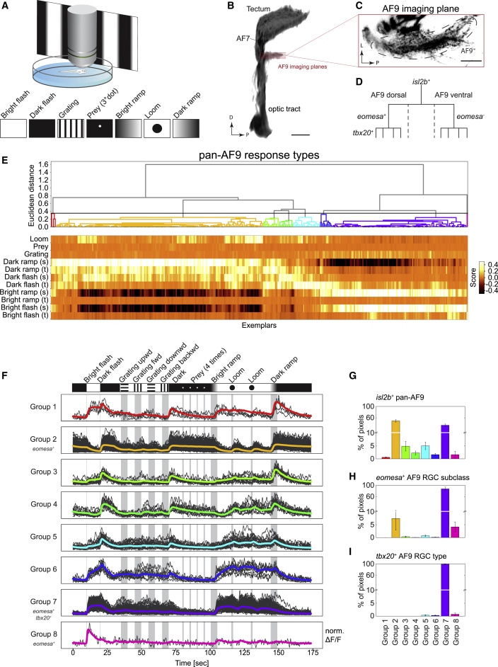
Fig. 6 (A) Functional imaging of RGC types. Neuronal activity was recorded using two-photon calcium imaging from immobilized larvae expressing GCaMP6s in RGC axon terminals during presentation of a battery of visual stimuli displayed on a projection screen. (B) 3D projection of the optic tract indicating the imaging planes in both ventral and dorsal subdivisions of AF9. D, dorsal; P, posterior. Scale bar, 50 μm. (C) Single imaging plane in AF9. L, lateral; P, posterior. Scale bar, 20 μm. (D) Hierarchical relationship of RGC subpopulations used for functional imaging: isl2b labels all RGCs, eomesa marks a subclass in dorsal AF9, wherein tbx20 is expressed by a single type among eomesa+ RGCs. (E) Diversity of isl2b+ RGC responses to visual stimuli in AF9-projecting axons. Neural activity recordings derived from single pixels were clustered using affinity propagation to reduce noise, resulting in 345 clusters represented by exemplars (STAR methods). Hierarchical clustering divided exemplar activity into eight major response groups (dendrogram, top). Heatmap (bottom) depicts calculated score of exemplars (columns) to each component of visual stimulus (rows). Sustained (s) and transient (t) activity was observed in responses to changing luminance levels. (F) Activity traces of eight classified response groups shown in (E) aligned with the visual stimulus sequence. Shown are the normalized averaged activity traces (colored lines) and all representing exemplars that fall into the group (gray lines) over time. Response groups encompass different numbers of exemplars depending on their abundance. (G–I) Relative frequencies of the eight response groups in isl2b+ RGCs (G), eomesa+ RGCs (H), and tbx20+ RGCs (I). Error bars represent SEM.