
|
Fig. S7
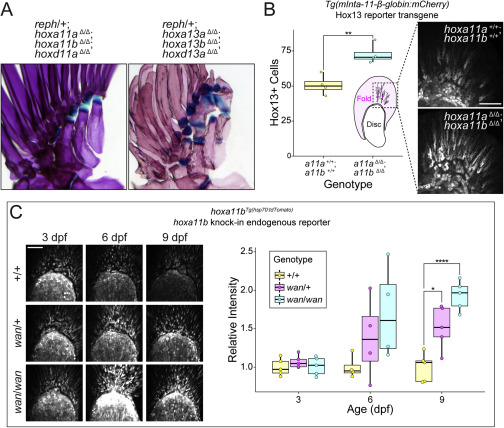
Figure S7. Interactions between Hox genes and reph and wan mutations, related to Figure 6 (A) Genetic interactions between reph and posterior Hox genes shape expression of the intermediate radial phenotype. reph heterozygotes that are triply homozygous null for hoxa11a, hoxa11b, and hoxd11a develop wild-type pectoral fin endoskeletons without intermediate radials. Heterozygous reph mutants triply homozygous for hoxa13a, hoxa13b, and hoxd13a exhibit an enhance endoskeleton elaboration phenotype, with each proximal radial producing intermediate radials. (B) HoxA11 paralogs negatively regulate Hox13 function in the fin. Double null mutants of hoxa11a and hoxa11b have significantly more Hox13-positive cells in the fin fold than do wild-type animals at 3 dpf (n = 4 each genotype, Welch’s Two Sample t test, t = 4.42, p = 0.0044). (C) hoxa11b expression is increased in wan mutants. Similar to reph mutants, wan mutants show higher levels of hoxa11b:tdTom expression in the early fin bud after 3 dpf. Intensity is significantly elevated in wan heterozygotes (single asterisk) and homozygotes (quadruple asterisk) at 9 dpf (n = 5 each genotype, Welch’s Two Sample t test, heterozygotes t = 3.43, p = 0.0055; homozygotes t = 7.82 p = 0.000026). Anterior to left, distal to top in all fin panels. Scale bars (B, C) 50 μm.
Reprinted from Cell, 184(4), Hawkins, M.B., Henke, K., Harris, M.P., Latent developmental potential to form limb-like skeletal structures in zebrafish, 899-911.e13, Copyright (2021) with permission from Elsevier. Full text @ Cell