
|
Fig. 2
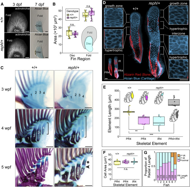
Figure 2. Novel intermediate radial elements arise by segmentation of a common precursor (A) The fin fold forms normally in reph/+ mutants. Immunolabeling of actinotrichia fibers at 3 dpf reveals no gross defects in the fin fold (n = 10). Alcian blue staining of cartilage at 7 dpf shows similar relative proportion of fin fold and endoskeletal disc size in wild-type and reph/+ animals. (B) Endoskeletal disc area at 7 dpf is not significantly different between wild-type and reph/+ fins (n = 18 each genotype, Welch’s Two Sample t test, t = −1.7605, p > 0.09). Fin fold area is also not significantly different (n = 18 each genotype, Welch’s Two Sample t test, t = 1.8918, p > 0.06). (C) Fin ontogeny in wild-type and reph/+ animals. Initial separation of the endoskeletal disc into the four proximal radial precursors is underway at 3 wpf. Proximal mineralization of the radials is evident at 4 wpf, while posterior radial cartilages are elongated in reph/+ mutants (bracket). At 5 wpf, wild-type proximal radials exhibit continuous zones of mineralization, while reph/+ mutants have a secondary bone collar (asterisk) separated from the proximal ossification by cartilage (arrowhead). (D) Elongated radials form interzones at sites of future segmentation. Confocal sections of wild-type and reph/+ radials at 5 wpf, bottom left inset shows full confocal projection, other insets magnify cartilage regions. (E) Quantification of radial element 4 lengths in wild-type and mutant animals at 5 wpf. Total reph/+ radials are significantly longer than wild-type PR4 (n = 7 each genotype, Welch’s Two Sample t test, t = 4.64, p = 8 × 10⁻⁴) while reph/+ PR4 elements are significantly shorter than wild-type elements (n = 7 each genotype, Welch’s Two Sample t test, t = −4.36, p = 1 × 10⁻³). (F) Quantification of average hypertrophic chondrocyte size at 5 wpf reveals no significant difference between wild-type and reph/+ elements (n = 7 each element type, one-way ANOVA, F = 0.319, p = 0.732). (G) Segmentation position along radial 4 is variable among reph/+ mutants. Absolute size of PR4 provided for each fish. Anterior to left, distal to top in all micrographs; PR4, proximal radial 4; IR4, intermediate radial 4; n.s., not significant; scale bars (A) 50 μm, (C) 125 μm, (D) 50 μm.
Reprinted from Cell, 184(4), Hawkins, M.B., Henke, K., Harris, M.P., Latent developmental potential to form limb-like skeletal structures in zebrafish, 899-911.e13, Copyright (2021) with permission from Elsevier. Full text @ Cell