
|
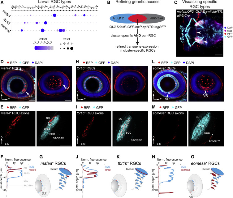
Fig. 4 (A) Dot plot showing expression patterns of mafaa, tbr1b, and eomesa (rows) in larval clusters (columns) ordered as in Figure 3A. Cluster numbers (top) correspond to immature (gray) and mature (black) RGC clusters. (B) Marker intersection refines genetic access to TF+ RGC types. In a TF:QF2 driver line, TF+ cells activate expression of GFP through a QUAS:switchNTR reporter. Combination with the pan-RGC Tg(ath5:Cre) line results in TF+ RGCs switching to RFP expression, while TF+ non-RGCs continue to express GFP. (C) Visualization of RGC types, shown here for mafaa+ RGCs (arrows, red labeling), by immunostaining in a triple-transgenic Tg(TF:QF2, QUAS:switchNTR, ath5:Cre) larva. Scale bar, 100 μm. (D–O) Anatomical characterization of RGC types labeled by mafaa (D–G), tbr1b (H–K), and eomesa (L–O) using quadruple-transgenic Tg(TF:QF2, QUAS:switchNTR, ath5:Cre, isl2b:GFP) larvae. In each case Tg(isl2b:GFP) serves as a label for landmarks of RGC projections. Confocal visualizations showing a single plane (left) and RGC soma distribution (maximum z projection, right) in en face views of the immunostained retina (D, H, and L), in vivo images of axonal projections in the tectum (E, I, and M), fluorescence profile across retinotectal laminae measured from the pan-RGC reporter isl2b and marker-specific RGC axons (F, J, and N) as well as a schematic representation of the soma distribution in the retina and the projection pattern indicating TF+ RGCs in red against all RGCs in blue (G, K, and O). Asterisks (∗) in (E, I, and M) denote layers innervated by TF+ RGCs. D, dorsal; T, temporal; A, anterior; M, medial. SZ, strike zone enrichment. VR, ventral retina enrichment. Scale bar in (D) for (D), (H), and (L), 50 μm. Scale bar in (E) for (E), (I), and (M), 50 μm.