
|
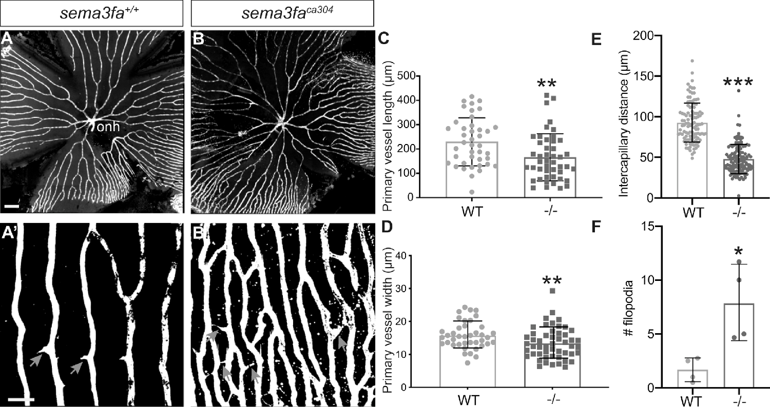
Fig. 3 Increased vascularization of the retina is observed into adulthood. (A, B) Vessels (white) in retinal flat mounts of Tg(kdrl:mCherry) WT siblings (A) and sema3faca304 (B) 5 month fish. Capillaries furthest from the optic nerve head (onh) in the peripheral retina are shown in A’,B’. A greater number of filopodial interconnections between capillaries are present in mutant retina (gray arrows). (C) Primary vessel length as measured from the optic nerve head (onh) to the first branching event is significantly shorter in mutants (**P = 0.0016, N = 2, n = 6 retinas) as compared to WT siblings (N = 2, n = 6 retinas). (D) The average width of the primary vessels is reduced significantly in mutants (**P = 0.0078; N = 2, n = 6 retinas) as compared to WT (N = 2, n = 6 retinas). (E) Measurement of the intercapillary distance shows that mutant capillaries are more closely spaced (n = 4 retinas, P < 0.0001) than in WT (n = 4 retinas). (F) Average number of filopodia in a region of interest in the retinal periphery (WT n = 4; sema3faca304 n = 4; P = 0.029). Error bars represent standard deviation (SD). Statistics represent the nonparametric Mann-Whitney U test. Scale bar: 200 µm (A) and 50 µm (A’).