
|
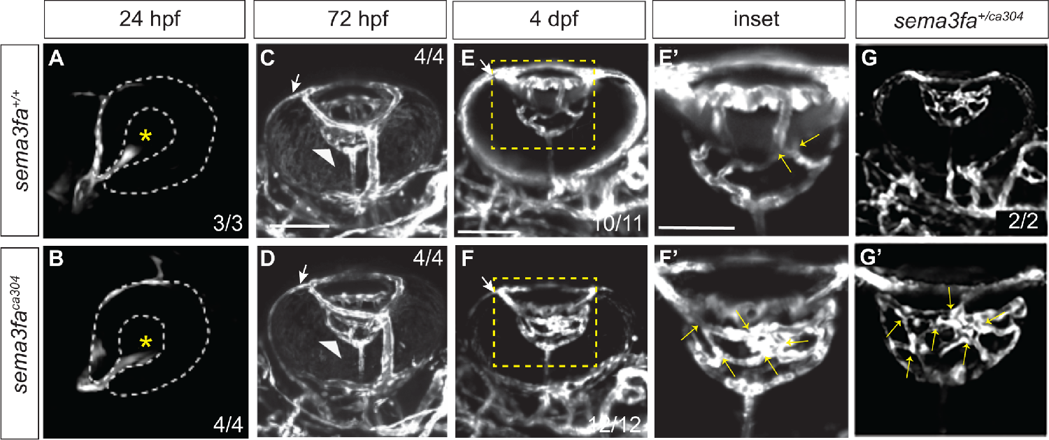
Fig. 2 Sema3fa is required to refine and limit retinal vessel branching in the larval eye. Live imaging of vessels (white) in Tg(kdrl:mCherry) WT (A,C,E,E’) and sema3faca304 heterozygous (G,G’), and homozygous (B,D,F,F’) eyes during retinal intraocular vascular development acquired by confocal microscopy. (A, B) In lateral views of WT (A) and sema3faca304 homozygous embryos, the hyaloid artery (endothelial cells labeled by mCherry) contacts the lens (asterisk) of the retina by 24 hpf (N = 1 WT n = 3; sema3faca304 n = 4). (C, D) Ventral views of WT (C: N = 1, n = 4) and sema3faca304 (D: N = 1, n = 4) eyes reveal that the hyaloid artery (arrowhead) forms a vascular plexus network around the lens by 72 hpf. Also evident is the nasal ciliary artery (arrows). (E–G) The WT intraocular hyaloid vessel (E’ is a magnified view of the boxed area in E) undergoes remodeling between 4-5 dpf to simplify the network (Hartsock et al., 2014). At 4 dpf, the numbers of vessel crossovers (arrows) are increased in sema3fa heterozygous (G,G’: N = 1, n = 2/2) and homozygous (F,F’: N = 4, n = 12/12) mutant embryos as compared to WT siblings (E, E’: N = 4, n = 10/11 show refinement). Scale bar: 100 µm; 50 µm for E’–G’.