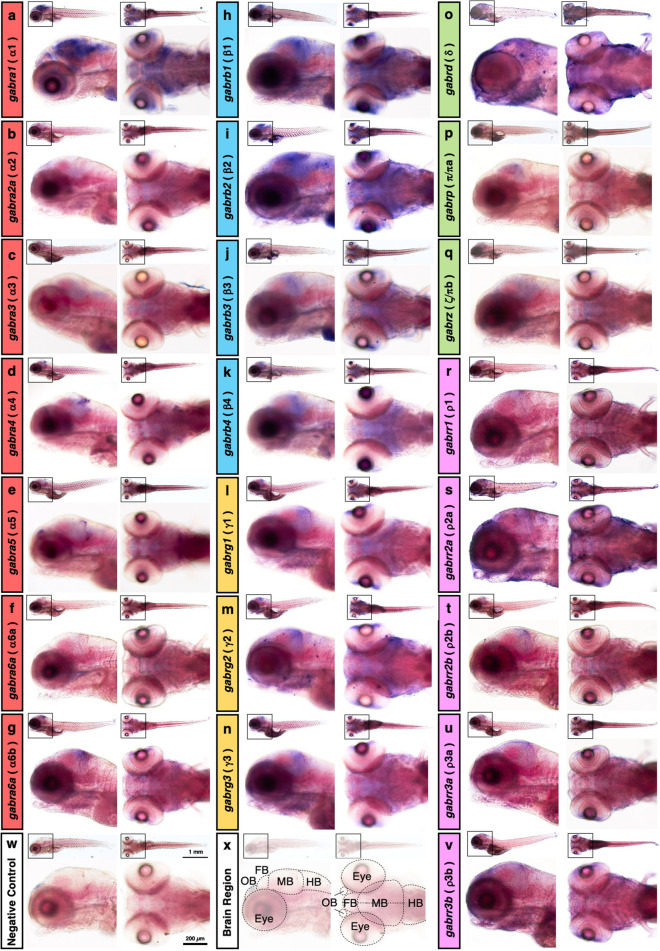
Image
Figure Caption

Figure 2

Spatial expression of zebrafish GABAA receptor subunits. Whole-mount in situ hybridization of 5 dpf zebrafish larvae using antisense probes for gabra1 (a), gabra2a (b), gabra3 (c), gabra4 (d), gabra5 (e), gabra6a (f), gabra6b (g), gabrb1 (h), gabrb2 (i), gabrb3 (j), gabrb4 (k), gabrg1 (l), gabrg2 (m), gabrg3 (n), gabrd (o), gabrp (p), gabrz (q), gabrr1 (r), gabrr2a (s), gabrr2b (t), gabrr3a (u), and gabrr3b (v). Negative control without probe showing no signals (w). Regions of the olfactory bulb, forebrain, midbrain, hindbrain, and eye are indicated in the image (x). Each labeling image is composed of a whole lateral view (left top), a whole dorsal view (right top), a magnified lateral view of the head region (left bottom), and a magnified dorsal view of the head region (right bottom). The scale bars in the whole and magnified views are 1 mm and 200 μm, respectively. OB: olfactory bulb; FB: forebrain; MB: midbrain; HB: hindbrain.

Figure Data
Acknowledgments
This image is the copyrighted work of the attributed author or publisher, and ZFIN has permission only to display this image to its users. Additional permissions should be obtained from the applicable author or publisher of the image. Full text @ Sci. Rep.