
|
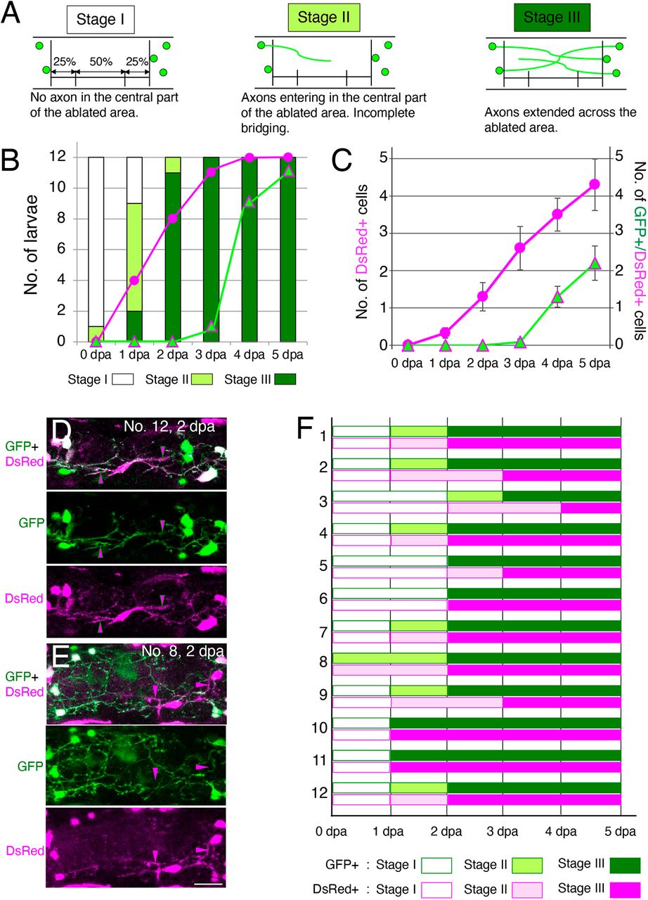
Fig. 5 Timecourses of the appearance of axons and ENCDCs in the ablated area during regeneration. (A) The appearance of neurites in the ablated area was classified into three stages (stages I-III) based on the degree of entry into ablated areas, as described below each schema. (B) The number of larvae at each stage for 12 operated larvae across the six timepoints examined (0 to 5 dpa). Magenta circles show the number of larvae with DsRed+ cells in the central 50% of the ablated area. Green triangles with magenta lines show the number of larvae with GFP+/DsRed+ cells in the same area. Neurite bridging was completed by 3 dpa for all operated larvae, and at that time, DsRed+ cells were also found in the central part of the ablated gut in 11 of the 12 larvae. GFP+/DsRed+ cells in the central part of the ablated area were found in most larvae (9/12) at 4 dpa. (C) The average number of DsRed+ and GFP+/DsRed+ cells in 12 operated larvae at the six time points examined (0 to 5 dpa). Magenta circles show the number of DsRed+ cells in the central 50% of the ablated area. Green triangles with magenta lines show the number of GFP+/DsRed+ cells in the same area. Data are presented as the mean±s.e.m. (D,E) Two examples of Red cells that entered into the ablated area. The Red cell in D extends its processes along the GFP+ processes (indicated by magenta arrowheads with green lines), whereas the Red cell(s) in E has/have DsRed+ processes extending independently of GFP+ neurites (magenta arrowheads). Scale bar: 25 µm. (F) Stages of GFP+ and DsRed+ processes at each day after ablation for 12 larvae (1-12). GFP+ and DsRed+ processes reached the same stage with the same timing for more than half (8/12) of larvae, while DsRed+ processes showed slight delay for four samples, suggesting there is no evidence that non-neuronal processes entered the ablated area to facilitate subsequent neurite extension.