
|
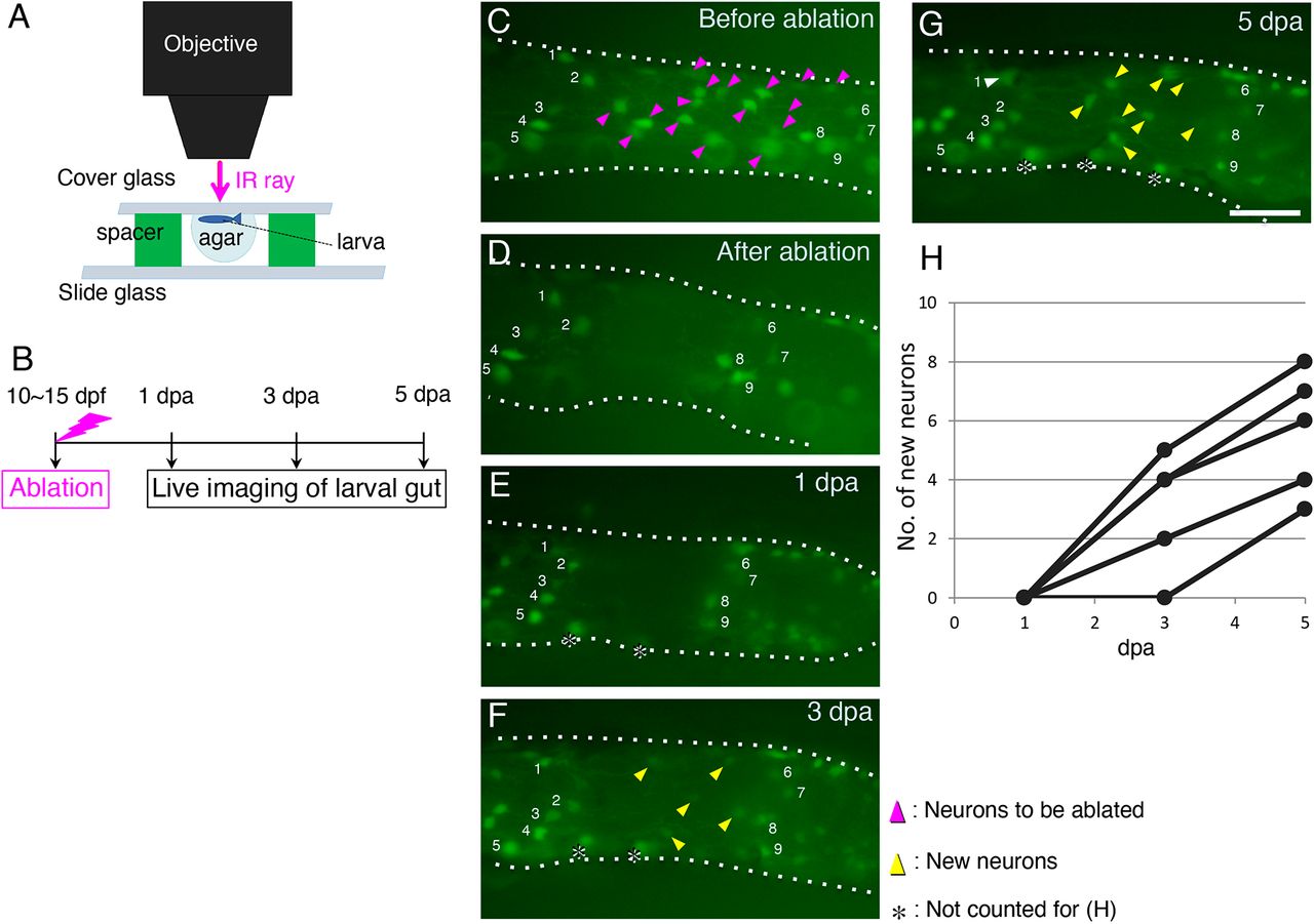
Fig. 2 Enteric neurons marked with GFP are regenerated after ablation. (A) Schematic of the ablation equipment. An anesthetized zebrafish larva of Tg(217B; u:gfp) (10-15 dpf) was mounted in agar under a coverglass with its left side facing up toward the objective. The neurons at the second-somite level from the anus were removed by irradiating an infrared (IR) ray through the objective (see Materials and Methods for details). (B) Timeline of the experiments. After ablation, each larva was cultured and live imaged at 1, 3 and 5 dpa. (C-G) Recovery of GFP+ cells after ablation in the same larva. Cells before ablation are marked with magenta arrowheads in the posterior intestine (C), and they were removed as shown in (D). Imaging at 1 dpa (E), 3 dpa (F) and 5 dpa (G) revealed new GFP+ cells (yellow arrowheads). Numbers in each panel mark identified cells before and after regeneration. Cells marked with asterisks were not counted for the data presented in H, because these cells are located near the ventral midline of the gut tube and are likely to have been hidden behind the gut when ablating. White arrowhead in G indicates the identified neuron 1. Intestines are outlined with dotted lines. Images are left side views of the distal intestine, with the anterior end positioned to the left of the image. Scale bar: 50 µm. (H) Numbers of new GFP+ cells in the ablated areas of five larvae examined at 1, 3 and 5 dpa during regeneration.