
|
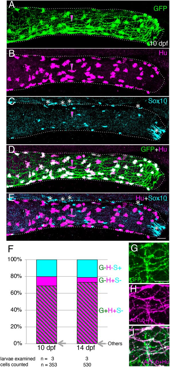
Fig. 1 Immunostaining of intestine in the transgenic line Tg(217B; u:gfp). (A-E) 10 dpf larval distal intestine stained for GFP (A), HuC/D (Hu, B) and Sox10 (C), with merge images shown (D,E). Dotted lines outline the intestine. Images are left side views, with the anterior end positioned to the left of the image. To show the overlapping expression of GFP and HuC/D, signal intensity for each marker is enhanced (A,B,D). Magenta arrowheads mark an example of a GFP−/Hu+ cell. The white asterisks in the most distal end of the intestine indicate unidentified Sox10 signals, and other asterisks around the dorsal part of intestine mark pigment cells outside of the intestine. Scale bar: 25 µm. (F) Proportions of GFP−/Hu−/Sox10+ (G−H−S+), GFP−/Hu+/ Sox10− (G−H+S−), GFP+/Hu+/Sox10− (G+H+S−) and other (Others) cell types at 10 and 14 dpf as a percentage of all cells examined (353 and 530 cells, respectively). ‘Others’ represent one GFP+/Hu+/Sox10+ and one GFP+/Hu−/Sox10+ cell at 10 dpf, and one GFP+/Hu+/Sox10+ cell at 14 dpf. Cells were counted in the distal part of the intestine, as exemplified in A-E. Numbers of examined larvae and cells at each stage are shown below the columns. (G-I) Co-expression of GFP and acetylated tublin (AcTub) in the distal intestine at 10 dpf. Markers shown are indicated in the bottom-left corners of panels. Images are left side views, with the anterior end positioned to the left of the image. Scale bar: 10 µm.