
Image
|
Figure Caption
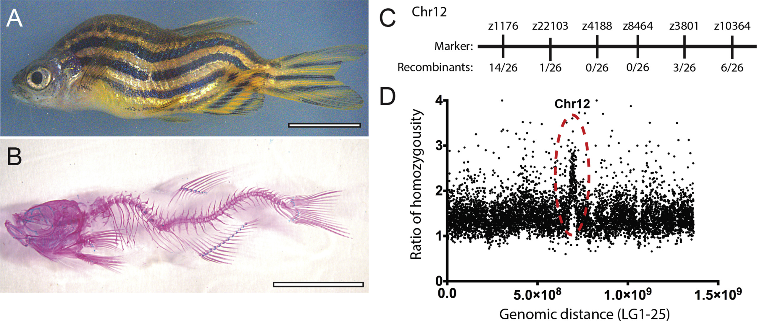
Fig. 7 The falkor scoliosis mutant. (A) Bright field and (B) Alizarin-Red/Alcian-Blue stained skeletal preparations illustrate the homozygous falkor mutant phenotype. (C) Meiotic mapping places the mutation on Chr. 12 in the neighborhood of z4188 and z3801. (D) Mapping by regions of homozygosity suggests a causative mutation in korken gene on Chr. 12.
Figure Data
Acknowledgments
This image is the copyrighted work of the attributed author or publisher, and
ZFIN has permission only to display this image to its users.
Additional permissions should be obtained from the applicable author or publisher of the image.
Reprinted from Developmental Biology, 471, Gray, R.S., Gonzalez, R., Ackerman, S.D., Minowa, R., Griest, J.F., Bayrak, M.N., Troutwine, B., Canter, S., Monk, K.R., Sepich, D.S., Solnica-Krezel, L., Postembryonic screen for mutations affecting spine development in zebrafish, 18-33, Copyright (2020) with permission from Elsevier. Full text @ Dev. Biol.