
Fig. 2
- ID
- ZDB-IMAGE-210316-28
- Publication
- Yu et al., 2020 - Taste buds are not derived from neural crest in mouse, chicken, and zebrafish
- All Figures
- Figures for Yu et al., 2020
|
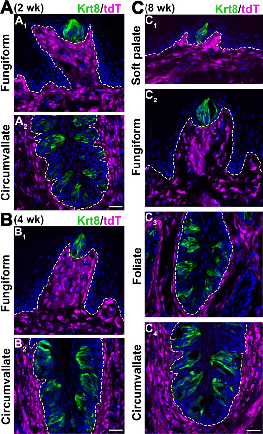
Fig. 2 Fig. 2. Single-plane laser scanning confocal photomicrographs to demonstrate the distributions of Sox10-iCreERT2/tdT-labeled cells in the tongue and soft palate in postnatal mice (TmxE7.5) at different stages. A-B: Images of a fungiform papilla on a sagittal section (A1, B1) and circumvallate on a coronal section (A2, B2) of tongue at 2 wk (A) and 4 wk (B). C: Images of soft palate (C1), fungiform papilla (C2), foliate papilla on a sagittal section (C3), and circumvallate papilla on a coronal section (C4) of tongue tissue at 8 wk. Taste buds were marked by the immunosignals of Keratin 8 (Krt8, green). White dashed lines demarcate lingual epithelium from the underlying connective tissue. Scale bars: 50 μm for all images.
Reprinted from Developmental Biology, 471, Yu, W., Wang, Z., Marshall, B., Yoshida, Y., Patel, R., Cui, X., Ball, R., Yin, L., Kawabata, F., Tabata, S., Chen, W., Kelsh, R.N., Lauderdale, J.D., Liu, H.X., Taste buds are not derived from neural crest in mouse, chicken, and zebrafish, 76-88, Copyright (2020) with permission from Elsevier. Full text @ Dev. Biol.