
|
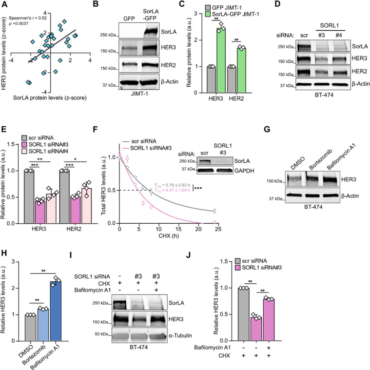
Fig. 3 A SorLA and HER3 protein levels correlate positively in breast cancer cell lines (DepMap portal; N = 29). B–E HER3 and HER2 expression correlates with SorLA in HER2-positive breast cancer. B, C SorLA-GFP transfection in JIMT-1 cells increases HER2 and HER3 levels compared to GFP transfected cells. D, E. SorLA silencing in BT-474 cells decreases HER2 and HER3. B, D Representative immunoblotting of total HER2, HER3, and SorLA with β-actin as a loading control. C, E represent the respective quantifications of immunoblots in (B, D) with HER2/HER3 levels normalized to loading control and relative to control-silenced cells. F SorLA silencing decreases HER3 stability. RNAi transfected BT-474 cells were treated with 25 µg.mL−1 of CHX for the indicated time points and HER3 protein levels were determined by immunoblotting (see Fig. S3E). Shown are HER3 levels normalized to α-tubulin and relative to 0 h timepoint. Half-lives (T1/2) represent the time required for HER3 to decrease to 50% of its initial level. The least squares fitting method and extra-sum-of-squares F test were used to assess the statistical difference between curves from control and SorLA-silenced cells (P = 0.0002). A representative western blot validating SorLA silencing is shown. G HER3 is primarily degraded through the lysosomal pathway. BT-474 cells were treated with 1 µM of bortezomib or 50 nM of bafilomycin A1 for 4 h to inhibit proteasome and lysosome activities, respectively. HER3 expression was analyzed by immunoblotting, with α-tubulin as a loading control. H Quantification of HER3 levels normalized to loading control and relative to DMSO-treated control cells. I SorLA silencing triggers HER3 lysosomal degradation. SorLA-silenced BT-474 cells were cotreated for 4 h with CHX and bafilomycin A1. HER3 expression was analyzed by immunoblotting, with α-tubulin as a loading control. J Quantification of HER3 levels normalized to loading control and relative to CHX-treated control-silenced cells. Data are mean ± SD from three (C, F, H, J) or four (E) independent biological replicates. Statistical analyses: Student’s t test (unpaired, two-tailed, unequal variance) unless indicated otherwise. Scr: control non-targeting siRNA.