IMAGE

Fig. 6

ID
ZDB-IMAGE-210307-71
Source
Figures for Póvoa et al., 2021
Image
Figure Caption

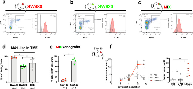
Fig. 6 Mice xenografts display similar TME behavior as zebrafish.

ac Representative graphs of flow cytometry analysis of the TME of SW480, SW620, and MIX mouse (Rag1−/−C57BL/6J) xenografts at 3 weeks post inoculation. d Quantification of double positive F4/80, CD80 macrophage in each TME, SW620 vs SW480. *P = 0.016, Cohen’s D g = 6.2; SW620 vs MIX ns = 0.29, Cohen’s D g = 0.83; SW480 vs MIX *P = 0.029, Cohen’s D g = 1.28). e Quantification of the percentage of each clone in MIX mice xenografts, *P = 0.029, Cohen’s D g = 7.68. d, e Data from quantification of flow cytometry analysis. Error bars represent mean ± S.D. f Growth curves of SW480 tumors treated with PBS, L-PBS, or L-Clodronate mice (f ns = 0.43, Cohen’s D g = 0.12, **P = 0.007, Cohen’s D g = 2.16, *P = 0.017, Cohen’s D g = 1.92). Error bars represent mean ± SEM. All data were analyzed using unpaired two-sided Mann–Whitney test. To avoid macrophage repopulation, mice were injected every 4 days (see “Methods”). N is depicted in the chart. Each dot represents one mouse xenograft. Source data are provided as a Source data file.

Acknowledgments
This image is the copyrighted work of the attributed author or publisher, and ZFIN has permission only to display this image to its users. Additional permissions should be obtained from the applicable author or publisher of the image. Full text @ Nat. Commun.