IMAGE

Figure 3

ID
ZDB-IMAGE-210305-11
Source
Figures for Lämmle et al., 2020
Image
Figure Caption

Figure 3

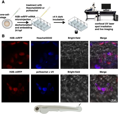
In vivo imaging of cell nuclei in zebrafish with pcHoechst. A) General schematic workflow for experiments in Figures 3, 4, S11, and S12. H2B‐RFP mRNA is injected into single‐cell‐stage zebrafish embryos for correlative nuclear staining of pcHoechst with a red fluorescent DNA‐associated fusion protein (Histone 2B fused to mRFP). Hoechst33342 or pcHoechst is applied (between 32–56 hpf), with 4–16 hours′ incubation in the dark before targeted uncaging and subsequent imaging. B) Agarose‐embedded H2B‐RFP‐expressing zebrafish embryos were incubated with 10 μM Hoechst 33342 (upper row) or 100 μM pcHoechst (lower row) overnight at 32 hpf. Spot irradiation using a 405 nm UV laser targeted at a large cluster of cells was performed for uncaging pcHoechst at 52 hpf. Confocal imaging of the targeted area at the zebrafish tail fin at 54 hpf shows colocalization of pcHoechst (blue, bottom row) with mRFP‐tagged H2B (red) in comparison to Hoechst33342 (blue, top row). Merge includes bright‐field image. The shown images are representative of three uncaging experiments using the same parameters.

Acknowledgments
This image is the copyrighted work of the attributed author or publisher, and ZFIN has permission only to display this image to its users. Additional permissions should be obtained from the applicable author or publisher of the image. Full text @ Chembiochem