
|
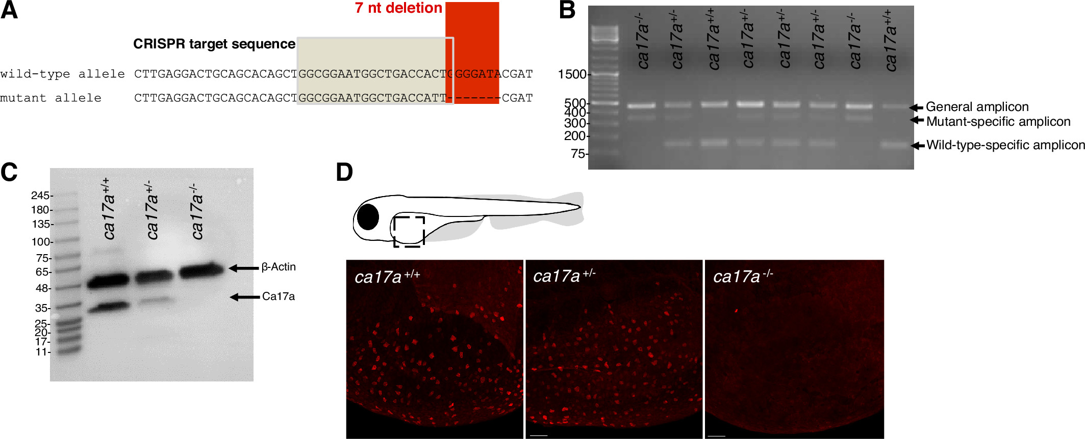
Fig. 1 Genotyping and confirmation of ca17a−/− deletion in larval zebrafish (Danio rerio). A: nucleotide alignment of the sequenced ca17a+/+ and ca17a−/− alleles showing the CRISPR sgRNA target sequence in the shaded box and the positions of the 7 nucleotide (nt) deletion and C to T substitution mutations in the red box. B: multiplex PCR products from fin clips of F2 larval offspring of F1 ca17a+/− crosses. The general amplicon (477 base pairs; bp) band appears for all genotypes, the 147-bp band is the ca17a+/+-specific amplicon, and the 352-bp band is the ca17a−/−-specific amplicon. C: representative Western blot of Ca17a and β-actin protein expression in ca17a+/+, ca17a+/−, and ca17a−/− whole larvae homogenates at 9 days postfertilization (dpf). D: immunofluorescent staining of Ca17a in the yolk sac of ca17a+/+, ca17a+/−, and ca17a−/− 4 dpf larval zebrafish. Scale bar = 50 μm.