
Image
|
Figure Caption
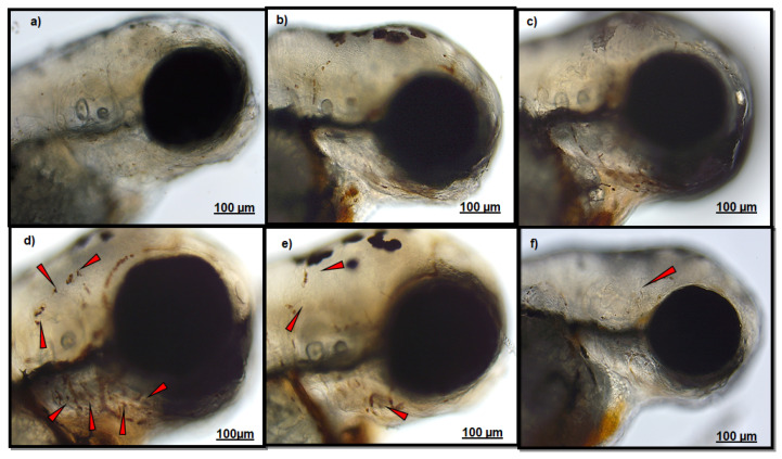
Figure 1 Figure shows representative images of neutrophils localization in the head of: (a) CTRL larvae; (b) polydatin (PD)-T0 larvae; (c) PD-T2 larvae; (d) CuSO4 larvae; (e) PD-CuSO4 – larvae; (f) CuSO4-PD larvae. Red arrows indicate neutrophil localization. Scale bar 100 µM.
Acknowledgments
This image is the copyrighted work of the attributed author or publisher, and
ZFIN has permission only to display this image to its users.
Additional permissions should be obtained from the applicable author or publisher of the image.
Full text @ Int. J. Environ. Res. Public Health