IMAGE

Fig. 2

ID
ZDB-IMAGE-210224-7
Source
Figures for Gerlach et al., 2021
Image
Figure Caption

Fig. 2

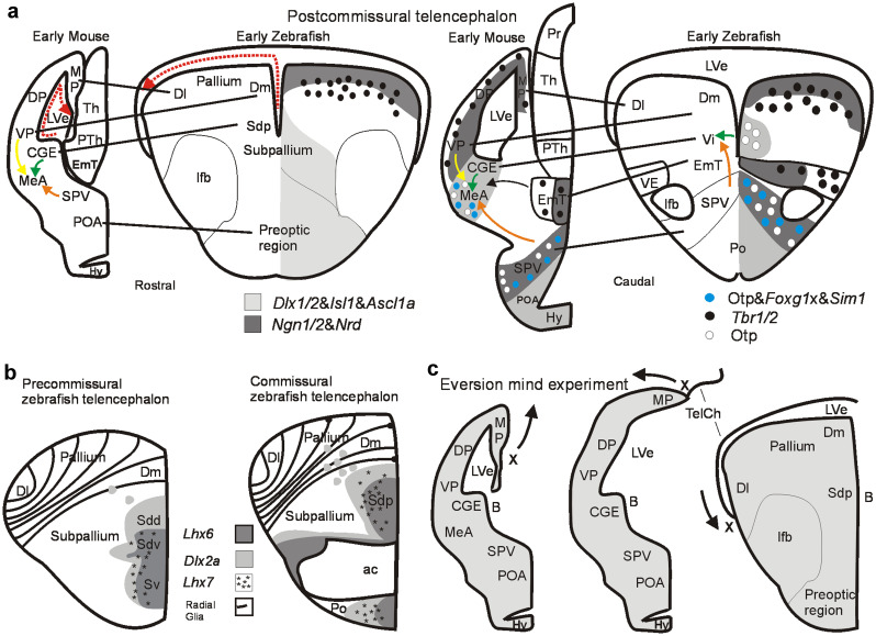
Development of the amygdala in early mouse and zebrafish brain. a Rostral (left panel) and caudal (right panel) transverse sections of postcommissural telencephalic mouse and zebrafish brains with critical pallial and subpallial gene markers indicated. Gene expression for mouse after Osório et al. 2010; Lo et al. 1991; Ma et al. 1997; Horton et al. 1999; Torii et al. 1999; Fode et al. 2000; Puelles et al. 2000), Morales et al. 2020; for zebrafish after Mueller et al. 2008; Herget et al. 2014, Affaticati et al. 2015; Mueller and Wullimann 2016 (see there for details). Dotted red arrows indicate topological lateromedial course of pallial ventricular surface. Solid yellow arrow indicates tangential migration of glutamatergic ventral pallial cells into mouse medial amygdala. Solid orange arrow indicates tangential migration of Otp positive glutamatergic cells from hypothalamic supraopto-paraventricular region (SPV) into medial amygdala (MeA; mouse)/intermediate nucleus of ventral telencephalon (Vi; zebrafish). Solid green arrow indicates radial migration of subpallial GABAergic cells into medial amygdala (mouse)/intermediate nucleus of ventral telencephalon (Vi, zebrafish). b Subpallial gene expression in early precommissural and commissural zebrafish telencephalon identifies within the larval subpallium a ventral division (Sv; adult ventral nucleus of ventral telencephalon, Vv, the septum homolog) and a dorsal division (Sd, adult dorsal nucleus of ventral telencephalon, Vd, the basal ganglia homolog). Sd is further genetically subdivided into ventral (Sdv) and dorsal (Sdd) subdivisions, representing the pallidal and striatal homologs, respectively (and embryonic mammalian medial and lateral ganglionic eminences). At the (commissural) level of the anterior commissure, the posterior division of the larval zebrafish dorsal subpallial division (Sdp) starts. In the adult zebrafish brain, it represents at this level the supracommissural nucleus of the ventral telencephalon (Vs). Panels in b modified after Mueller et al. (2008). The larval Sdp continues into postcommissural levels (seen in a left panel), represented in the adult zebrafish brain by the postcommissural nucleus of the ventral telencephalon (Vp). Only recently, a most posterior ventral telencephalic (subpallial) area characterized by otp positive cells was recognized (Herget et al. 2014; Biechl et al. 2017) and identified as the intermediate nucleus of the ventral telencephalon (Vi) in larval and adult zebrafish brains (see a, right panel, and Fig. 1). Note that the course of radial glia fibers is indicated in the zebrafish pallium. c This mind experiment illustrates how a postcommissural early rodent telencephalon is transformed into a postcommissural early zebrafish telencephalon by everting pallial masses. The most medial rodent pallial division (MP/hippocampus; note position of letter X and arrow indicating eversion) is virtually rolled out laterally resulting in the topography of the postcommissural teleostean/zebrafish telencephalon by maintaining topological relationships of major telencephalic divisions (see text). Compare a similar figure in Wullimann (2009) for the precommissural telencephalon. Abbreviations: ac, anterior commissure; CGE, caudal ganglion eminence; Dl, lateral zone of dorsal telencephalic area; Dm, medial zone of dorsal telencephalic area; DP, dorsal pallium; EmT, eminentia thalami; Hy, hypothalamus; lfb, lateral forebrain bundle; LVe, lateral ventricle; MeA, medial amygdala; MP, medial pallium; Po, preoptic area (zebrafish); POA, anterior preoptic area (mouse); PTh, prethalamus; Sd larval dorsal part of subpallium; Sdd, dorsal subdivision of Sd (striatum homomog); Sdv, ventral subdivision of Sd (pallidum homolog), Sdp, posterior subdivision of Sd (subpallial amygdala homolog); SPV, supraopto-paraventricular region; Sv, larval ventral part of subpallium; TelCh, tela chorioidea, Th, (dorsal) thalamus; VE, ventral entopeduncular nucleus; Vi, intermediate nucleus of ventral telencephalon; VP, ventral pallium

Acknowledgments
This image is the copyrighted work of the attributed author or publisher, and ZFIN has permission only to display this image to its users. Additional permissions should be obtained from the applicable author or publisher of the image. Full text @ Cell Tissue Res.