
Fig. 2
|
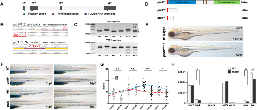
Fig. 2 Fig. 2. Lack of pax9 selectively abrogates neutrophils. (A) Structure of pax9 locus in zebrafish. Second exon was targeted by gRNA to generate pax9 knockout mutants. (B) Nucleotide sequence of pax9 locus was affected by two independent mutations. (C) The presence of mutation was validated by PCR-gel electrophoresis combination (top) and heteroduplex analyses using T7 endonuclease 1 (bottom). (D) In silico protein structure prediction of two mutants showing signaling peptide (Red), paired-box domain (Blue), and trans-activating domain (Green). (E) Gross morphology of 96hpf pax9 mutants. (F) The number of neutrophils was assessed by Sudan black staining. (G) Quantification of (F). Blue and Red dashes represent dynamic changes of neutrophil numbers in WT and mutant, respectively. dpf; day post fertilization (H) The expression of neutrophil specific markers was significantly reduced in 1.5dpf pax9 mutants compared to WT while other hematopoietic markers show no change except cmyb. . (For interpretation of the references to colour in this figure legend, the reader is referred to the Web version of this article.)