IMAGE

FIGURE 3

ID
ZDB-IMAGE-210221-3
Source
Figures for Lahne et al., 2021
Image
Figure Caption

FIGURE 3

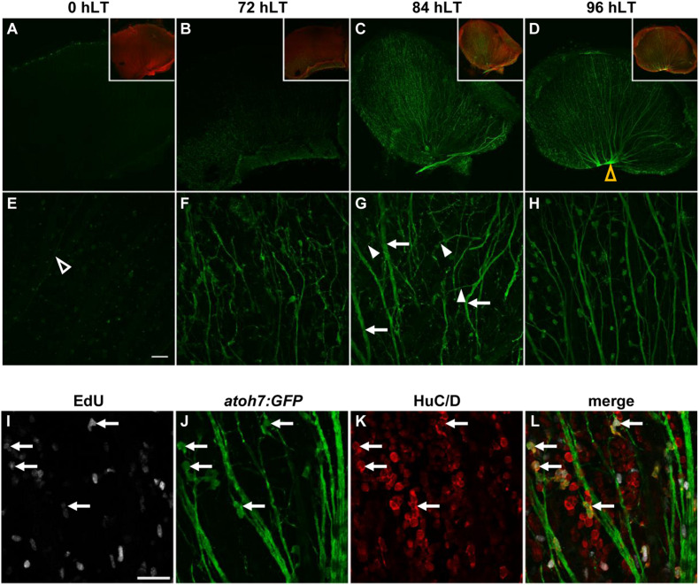
Newly generated ganglion cells extend axons. (A–H) Confocal images of dorsal retinal flatmounts from light-damaged Tg[atoh7:GFP]rw021 zebrafish (0, 72, 84, 96 hLT) immunolabeled for GFP (A–H) and phosphorylated gap43 to identify the nerve fiber layer (A–D, inset) at lower (A–D, single z-plane) and higher magnification [(E–H); maximum projections of five z-levels of the GCL]. Open yellow arrowhead, optic nerve head (D). Open white arrowhead, thin axonal projection (E). Filled arrowheads, neurites extending at an angle relative to thickened axon tracks (arrows). Scale bar, 50 μm (E). (I–L) Single z-plane confocal images of an EdU-injected (I,L)Tg[atoh7:GFP]rw021 dorsal retinal flatmount at 96 hLT immunolabeled for GFP (J,L) and HuC/D (K,L). Arrows indicate newly generated ganglion or amacrine cells. Scale bar, 20 μm (I).

Acknowledgments
This image is the copyrighted work of the attributed author or publisher, and ZFIN has permission only to display this image to its users. Additional permissions should be obtained from the applicable author or publisher of the image. Full text @ Front Cell Dev Biol