
|
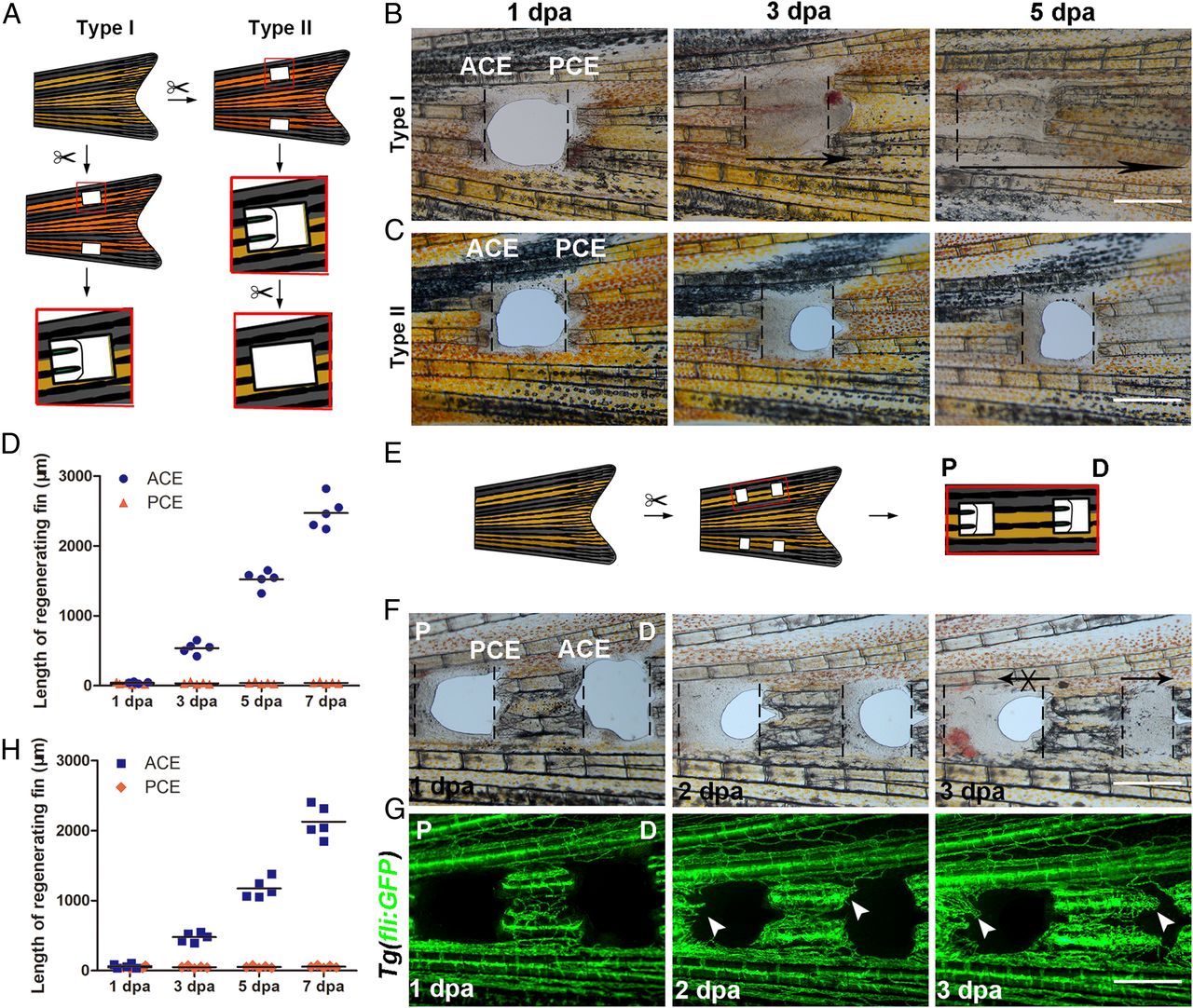
Fig. 1 P–D regeneration polarity occurrs in zebrafish fins. (A) Schematic of two types of fin excavation. (B and C) Type I (n = 11/11) and II (n = 11/12) experimental strategies of unidirectional regeneration in zebrafish fins from ACE to the PCE. (D) Quantification of fin regenerative tissue lengths (in micrometers) of the ACE and PCE (n = 5). (E) Schematic of the two-hole excavation in the same two bony rays. (F and G) Images of bright fields and blood vessels in fin regeneration after two-hole excavations at 1, 2, and 3 dpa (n = 5/8). (H) Quantification of fin regenerative tissue lengths (in micrometers) with two-hole excavation (n = 5). P, proximal; D, distal. (Scale bar: 500 µM.)