
Fig. 3
- ID
- ZDB-IMAGE-210218-56
- Publication
- Kim et al., 2020 - Notch Signaling Controls Oligodendrocyte Regeneration in the Injured Telencephalon of Adult Zebrafish
- All Figures
- Figures for Kim et al., 2020
|
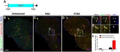
Fig. 3 Fig. 3. Regeneration of mature oligodendrocytes in the injured telencephalon. All panels show transverse sections of the telencephalon of adult zebrafish, with the dorsal side at the top. (A) Scheme of BrdU treatment. (B~D) Labeling of the telencephalon of Tg(mbp:EGFP) adult zebrafish with anti-BrdU antibody. Yellow arrows indicate the LVZ area of the control (B) and injured hemispheres of the telencephalon (C, D). (C’, D’) Boxed areas in C and D indicate the parenchymal area near the injury site. Arrows indicate mbp:EGFP+/BrdU- pre-existing mature oligodendrocytes (C’), and arrowheads indicate mbp:EGFP+/BrdU+ newly generated mature oligodendrocytes (D’). (E) Quantification of newly generated EGFP+/Dsred2+/BrdU+ mature oligodendrocytes (n=7 sections from two zebrafish, Mann–Whitney U test, *p<0.05). Scale bars: B~D, 100 μm; C’~D’, 25 μm. BrdU, bromodeoxyuridine; EGFP, enhanced green fluorescent protein; LVZ, lateral ventricular zone.