
|
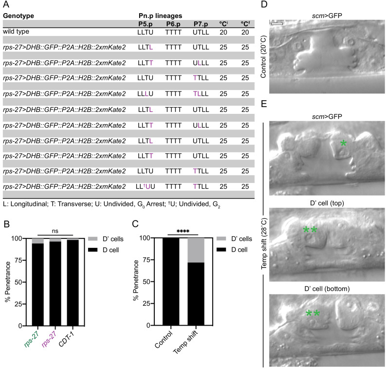
Figure 5—figure supplement 1. Related to Figure 5. (A) Pn.p lineages for respective genotypes listed. Letters indicate whether and in which orientation the four granddaughters further divided, following the nomenclature of Sternberg and Horvitz, 1986. Purple text indicates deviation from wild type condition. (B, C) Bar graphs displaying penetrance of D’ division in animals grown at 25 °C expressing different variants of the CDK sensor or endogenously tagged CDT-1::ZF::GFP or from temperature shifts from 20–28°C (C). (D, E) Representative DIC micrographs of 20 °C control (D) and 28 °C experimental (E) conditions of L4 stage vulva with an extra D cell (D’) division (**) as compared to wild type (*). Scale bar = 5 µm.