
|
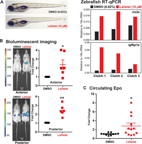
Fig. 6 Figure 6. LAL inhibition activates HIFα in animals. A, representative images of WT zebrafish larvae (5 days postfertilization) treated with lalistat (10 μm) or vehicle control DMSO (0.02%) for 2 h are shown. Expression of HIFα targets irs2a and igfbp1a (relative to 18s rRNA, RNA pooled from five larvae) was analyzed using RT-qPCR. B, ODD-Luc mice received subcutaneous injection of either DMSO or lalistat (20 mg/kg) three times per week for 2 weeks. Shown are representative bioluminescent images (captured 2 min after luciferin injection) of ODD-Luc mice treated as indicated. Bioluminescent signals from lalistat-treated mice were normalized to vehicle control (n = 6, mean ± S.E. (error bars)). p values from a single-column t test (Lalistat versus DMSO) are shown: *, p < 0.05; **, p < 0.005. C, circulating Epo levels from lalistat-treated mice (n = 12, mean ± S.E.) were normalized to vehicle control (n = 10, mean ± S.E.). p values from a single-column t test (lalistat versus DMSO) are shown: *, p < 0.05.