
|
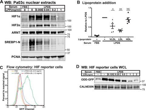
Fig. 2 Figure 2. Low-density lipoprotein regulates HIFα. A, immunoblots (WB) of nuclear extracts from Pa03c cells cultured for 16 h in FBS or LPDS supplemented with bovine lipoproteins (LPP; 0–1 mg/ml). PCNA served as a loading control. B, Pa03c cells were cultured for 16 h in FBS or LPDS with the indicated lipoprotein additions: human VLDL (0.2 mg/ml), human LDL (1 mg/ml), and human HDL (0.5 mg/ml). HIF1α immunoblot signal was normalized first to the loading control HDAC1 and then normalized to that in LPDS (n = 5, mean ± S.E. (error bars)). p values from a single-column t test (LPDS versus LPDS + VLDL, LDL, or HDL) are shown; NS, not significant; *, p < 0.05; **, p < 0.005. C, flow cytometry analysis of HIF reporter cells cultured for 24 h in FBS, LPDS, or LPDS supplemented with 0.5 or 1.5 mg/ml human LDL. D, immunoblots of whole-cell lysates from HIF reporter cells cultured for 24 h in FBS, LPDS, or LPDS with the indicated concentrations of human LDL. CALNEXIN served as a loading control.