
|
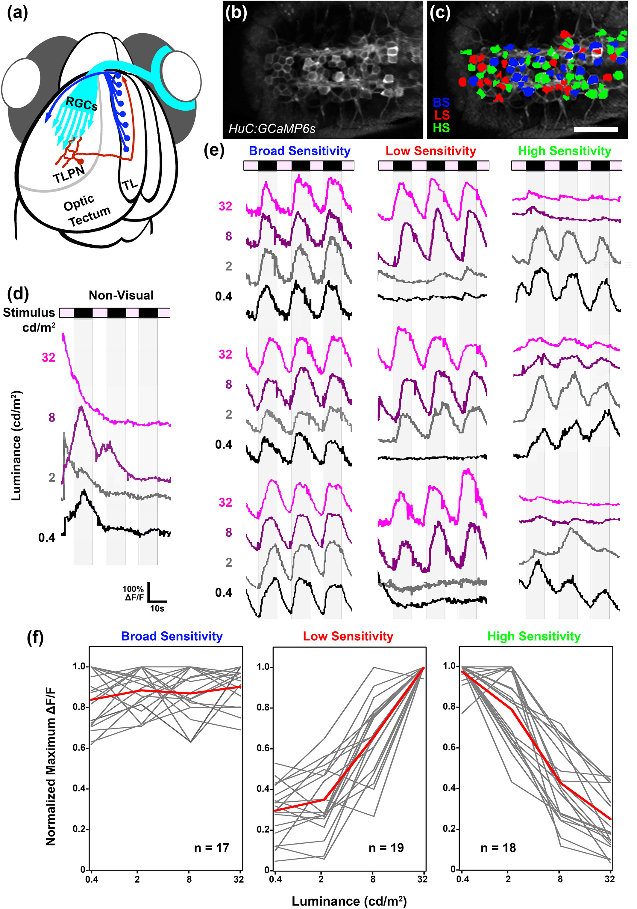
Fig. 8 Parallel dimming pathways remain segregated in torus longitudinalis (TL). (a) Schematic of retinal ganglion cell innervation of the tectum and reciprocal connection between the tectum and TL. (b) Single multiphoton image from a recording in the TL of a 6 dpf Tg(elavl3:gal4,uas:gcamp6s) larva presented with light steps to the right eye. Anterior is to the right. (c) Region of interest color coding indicates photosensitivity profile of individual TL neurons: broad sensitivity (BS; blue), low sensitivity (LS; red), and high sensitivity (HS; green). Note dense activation of many neurons on both sides of the TL. Anterior is to the right. (d) Sample trace of a nonvisual TL neuron. Note large events that are highly variable with respect to stimulus timing across trials. (e) Sample traces of individual BS, LS, and HS TL neurons. Each trace is 58 s in duration and consists of four ON phases of 7‐s duration interleaved with three OFF phases of 10‐s duration. Note that in several traces, baseline drifted in either the positive or negative direction. (f) Photosensitivity tuning curves for 54 dimming‐responsive neurons detected in TL (gray). Average of individual traces is shown in red. Scale bar: 40 μm [Color figure can be viewed at