
|
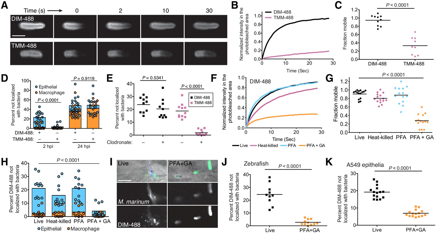
Fig. 5 PDIM’s mobility promotes spread into epithelial cell membranes. (A) Representative FRAP images of DIM-488 and TMM-488 labeled M. marinum, scale bar = 2 μm. (B) Fluorescent recovery curves after photobleaching of DIM-488 or TMM-488 labeled M. marinum, lines represent the average signal from n = 10 cells. (C) Mean fraction mobile which is the plateau following fitting of data generated in B to a non-linear regression with a one-phase association. (D) Mean percent DIM-488 or TMM-488 in macrophage or epithelial cells not localized with bacteria following HBV infection with ~100 M. marinum. (E) Mean percent DIM-488 or TMM-488 not localized with bacteria 24 hr following HBV infection of lipo-PBS or lipo-clodronate treated fish with ~100 M. marinum. (F) Fluorescent recovery curves after photobleaching of live, heat-killed, 4% paraformaldehyde (PFA) fixed, or 4% paraformaldehyde plus 1% glutaraldehyde (PFA+GA) fixed DIM-488 labeled M. marinum, lines represent the average signal from n = 14–15 cells. (G) Mean fraction mobile which is the plateau following fitting of data generated in F to a non-linear regression with a one-phase association. (H) Mean percent DIM-488 in macrophage or epithelial cells not localized with bacteria 2 hr following HBV infection with ~100 M. marinum treated as in F. (I) Images of live or PFA+GA treated DIM-488 labeled M. marinum at 2 hpi of the HBV with ~100 bacteria, scale bar = 5 μm. Mean percent DIM-488 not localized with bacteria 24 hr following (J) infection of lipo-clodronate treated fish or (K) A549 epithelial cells with live or PFA+GA fixed DIM-488 labeled M. marinum. (C), (J), and (K) two-tailed, unpaired t test. (E), (G), and (H) ordinary one-way ANOVA with Tukey’s multiple comparisons test with selected adjusted P values shown. (B)-(H) and (J)-(K) representative of three separate experiments.