IMAGE

Figure 5; supplement 3.

ID
ZDB-IMAGE-210208-77
Source
Figures for Alyenbaawi et al., 2021
Image
Figure Caption

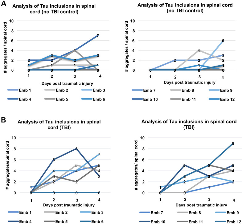
Figure 5; supplement 3. Longitudinal analysis of individual fish after traumatic brain injury (TBI) shows various patterns of Tau inclusion formation and clearance in their spinal cords.

(A) Exemplar data from the control group (no TBI) wherein many of the Tau4R-GFP biosensor larvae did not develop aggregates in the spinal cord, while a few either developed aggregates at later timepoints, or developed aggregates that disappeared later on. (B) Tau4R-GFP biosensor larvae subjected to TBI developed more GFP+ spinal cord aggregates and at earlier timepoints compared to controls, and the number of aggregates typically (although not always) increased with time. Within panels A and B, the left and right graphs are equivalent and display several larvae each.

Acknowledgments
This image is the copyrighted work of the attributed author or publisher, and ZFIN has permission only to display this image to its users. Additional permissions should be obtained from the applicable author or publisher of the image. Full text @ Elife