
|
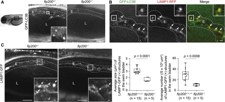
Fig. 2 Figure 2. Defective Maturation of Lamellar Bodies in Swim Bladder Epithelial Cells in fip200-Deficient Zebrafish (A) Representative images of GFP-LC3B signals in swim bladders of 4-dpf fip200+/− and fip200−/− zebrafish expressing GFP-LC3B. Scale bar, 20 μm and 1 μm in the inset. (B) Representative images of GFP-LC3B and LAMP1-RFP (lamellar bodies and endolysosomes) signals in swim bladders of 4-dpf wild-type zebrafish expressing GFP-LC3B and LAMP1-RFP. Arrows indicate LC3B- and LAMP1-positive structures. Scale bar, 2 μm. (C) Representative images of LAMP1-GFP signals in swim bladders of 4-dpf fip200+/− and fip200−/− zebrafish expressing LAMP1-GFP. Arrows indicate LAMP1-positive structures. Scale bar, 20 μm and 1 μm in the inset. The average size and number of lamellar bodies were quantified and are shown in the graphs. The solid bars and boxes indicate the median and interquartile range (25th to 75th percentile), respectively. The whiskers indicate the largest and smallest values. Differences were statistically analyzed by unpaired two-tailed Mann-Whitney U tests. E, epithelial cells; L, lumen of the swim bladder.